Download
1 / 38
390 likes | 440 Views
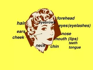
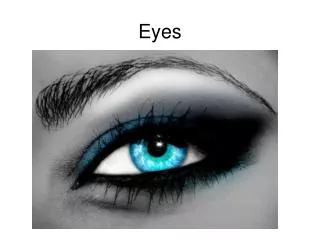
Eyes. 3.03 Remember the structures of the sensory system. 1. Warm up. Read pages in textbook. Fill out handout. Structures of the eyes. External structures Orbit Eyelids Eyelashes Conjunctiva Lacrimal apparatus Extrinsic muscles. 3.03 Remember the structures of the sensory system. 3.

E N D
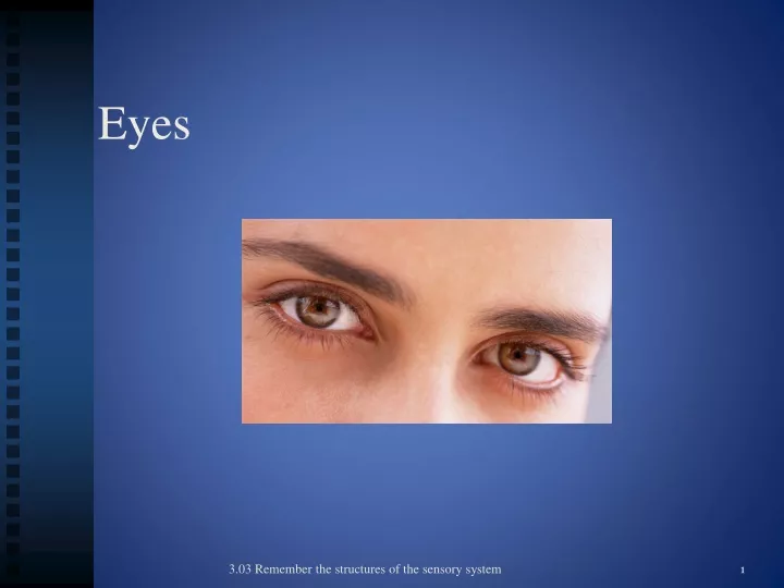
Eyes 3.03 Remember the structures of the sensory system 1
Warm up Read pages in textbook. Fill out handout. 3.04 Understand the functions and disorders of the sensory system
Structures of the eyes • External structures • Orbit • Eyelids • Eyelashes • Conjunctiva • Lacrimal apparatus • Extrinsic muscles 3.03 Remember the structures of the sensory system 3
Structures of the eyes • 1” in diameter • Protected by orbital socket of skull, eyebrows, eyelashes and eyelids • Bathed in fluid from LACRIMAL GLANDS – tears empty into nasal cavity • CONJUNCTIVA – thin membrane that lines the eyelids and covers part of the eye, secretes mucous to lubricate eye • Wall of the eye made up of three coats (sclera, choroid coat, retina) Two Chambers • Anterior containing iris, ciliary body and lens • Posterior- retina, macula, optic nerve. 3.03 Remember the structures of the sensory system 4
Eye Interesting tidbit External eye • Orbit • Eyelids and eyelashes Women blink twice as often as men. Why do we blink? • Conjunctiva • Lacrimal apparatus • Extrinsic muscles 3.04 Understand the functions and disorders of the sensory system
Eye Which extrinsic muscle allows you to look upward? External eye Extrinsic muscles- 4 Superior rectus, Inferior rectus, Medial rectus and Lateral rectus 3.04 Understand the functions and disorders of the sensory system
Eye Internal eye Sclera • Outer layer • White of the eye • Tough coating, helps maintain shape of eye and protects what’s inside • Muscles responsible for moving the eye are attached to the sclera – called EXTRINSIC MUSCLES 3.04 Understand the functions and disorders of the sensory system
Eye Internal eye • Cornea- the transparent front part of the eye that covers the iris, pupil, and anterior chamber Refracts light. • Choroid Coat- Layer of the sclera that contains the blood vessels-Blood supply to the eye 3.04 Understand the functions and disorders of the sensory system
Eye Internal eye Iris- thin, circular structure in the eye, responsible for controlling the diameter and size of the pupil-the amount of light reaching the retina. The color of the iris is often referred to as “ eye color “ Pupil a hole located in the center of the iris of the eye that allows light to enter the retina 3.04 Understand the functions and disorders of the sensory system 9
Eye Internal eye • Ciliary body- Muscles inside the eye -has three functions: accommodation, aqueous humor production and the production and maintenance of the lens zonules • Lens- a nearly transparent biconvex structure suspended behind the iris of the eye, the sole function of which is to focus light rays onto the retina 3.04 Understand the functions and disorders of the sensory system
Eye Internal eye Aqueous humor clear, watery fluid circulating in the chamber of the eye between the cornea and the lens. - nourishes the cornea and the lens and gives the eye it's shape. 3.04 Understand the functions and disorders of the sensory system 11
Eye Internal eye Vitreous humor- clear gel that fills the space between the lens and the retina. Helps keep the retina in place 3.04 Understand the functions and disorders of the sensory system 12
Eye Internal eye • CHOROID COAT • Middle layer • Blood Supply to retina • Opening in front is the PUPIL • Colored, muscular layer surrounding pupil is IRIS • INTRINSIC MUSCLES – change size of iris to control amount of light entering through the pupil 3.04 Understand the functions and disorders of the sensory system 13
Eye Internal eye RETINA Innermost layer Light rays focus an image on the retina The image travels to the cerebral cortex via the OPTIC NERVE Receives light focused from the lens, convert the light into neural signals, and send these signals on to the brain. 3.04 Understand the functions and disorders of the sensory system 14
Eyes • Rods and cones- specialized nerve fiber in the retina. • Rods- Located on the pouter edges of the retina • Do not perceive color • Function in dim light allowing us to see and also helps with peripheral vision • Cones • Active in bright light and allow you to precieve color • If you don’t have cones you are considered color blind. 3.03 Remember the structures of the sensory system 15
Eye Internal eye • OPTIC DISC – Located on the retina on the back of the eye. • Known as the blind spot – nerve fibers gather here to form the optic nerve, no rods or cones 1 Million Neurons 3.04 Understand the functions and disorders of the sensory system 16
a small and highly sensitive part of the retina responsible for detailed central vision. Eye What is the macula? 3.04 Understand the functions and disorders of the sensory system
Eye Why can’t you see in the dark? Process of Seeing • Trace the field of vision. • Light • Cornea • Pupil • Lens (where light rays are refracted) • Retina • Rods and Cones (pick up stimulus) • Optic Nerve • Brain • Is there anything strange about this picture? Explain 3.04 Understand the functions and disorders of the sensory system
Eye Vision What happens as you move your paper away from and toward to your eye? 3.04 Understand the functions and disorders of the sensory system
Eyes Name the structures… • Review 20 3.03 Remember the structures of the sensory system
Check your knowledge! 3.04 Understand the functions and disorders of the sensory system
http://www.illusions.org/ Vision Activity Do you see what I see? 3.04 Understand the functions and disorders of the sensory system
Disorders of the eye Astigmatism Presbyopia Glaucoma Diabeticretinopathy Detachedretina Hyperopia Color blindness Cataract Myopia Conjunctivitis Have you heard of these conditions? What do you know about them? 3.04 Understand the functions and disorders of the sensory system
Disorders of the eye Cataract Describe this lens. 3.04 Understand the functions and disorders of the sensory system
Disorders of the eye Cataract How is a cataract treated? 3.04 Understand the functions and disorders of the sensory system
Disorders of the eye • CATARACTS • Lens of eye gradually becomes cloudy • Frequently occurs in people over 70 • Causes a gradual blurring and loss of vision • Pupil turns from black to milky white • Rx – surgical removal of the lens 3.04 Understand the functions and disorders of the sensory system
Disorders of the eye Ishihara chart Color blindness Do you see the number? Unable to distinquish between the colors red and green. Will most often only be able to see black, white and gray. A decrease in the number of rods in the eye. Men are more likely to be color blind 3.04 Understand the functions and disorders of the sensory system
Disorders of the eye How is conjunctivitis spread? How can it be prevented? Conjunctivitis Pink eye Inflammation of conjunctival membranes in front of the eye Redness, pain, swelling and discharge Highly contagious Rx – antibiotic eye drops 3.04 Understand the functions and disorders of the sensory system
Detached retina Discuss what happens in this process. What is the relevance to health? 3.04 Understand the functions and disorders of the sensory system
Detached retina Compare this process to the previous picture. What might cause this condition? 3.04 Understand the functions and disorders of the sensory system
Disorders of the eye Diabetic retinopathy • What causes diabetic retinopathy? • What are the symptoms? • Explain the impact on vision. 3.04 Understand the functions and disorders of the sensory system
Disorders of the eye Glaucoma • Excessive intraocular pressure causing destruction of the retina and atrophy of the optic nerve • Caused by overproduction of aqueous humor, lack of drainage, or aging • Sx – develop gradually – mild aching, loss of peripheral vision, halo around the light • TONOMETER – measures intraocular pressure • Rx – drugs or laser surgery 3.04 Understand the functions and disorders of the sensory system
Disorders of the eye Macular degeneration What is macular degeneration? Compare the two types. How is it diagnosed? What is the treatment? 3.04 Understand the functions and disorders of the sensory system
Problems with Focus 3.04 Understand the functions and disorders of the sensory system
PRESBYOPIA Lens loses elasticity, can’t focus on close or distant objects Usually occurs after age 40 Rx – Bifocals HYPEROPIA Farsighted Focal point beyond the retina because eyeball too short Convex lenses help 3.04 Understand the functions and disorders of the sensory system
MYOPIA Nearsighted Eyeball too long Concave lenses help ASTIGMATISM Irregular curvature of the cornea or lens, causing blurred vision and eye strain Rx – corrective lenses 3.04 Understand the functions and disorders of the sensory system
Testing vision SNELLEN EYE CHART – chart that uses letters or symbols in calibrated heights to check for vision defects 3.04 Understand the functions and disorders of the sensory system
Processing Labeling Activity 1 Look into my eyes