Download
1 / 44
450 likes | 582 Views
3- and 4-Dimensional Ultrasound Imaging in Obstetrics. Curtis Lowery, M.D. Professor and Section Head of MFM Medical Director of ANGELS. 2D to 3D. The mental process of converting 2D into 3D images is not an easy one, and is dependent on individual skills and training.

E N D
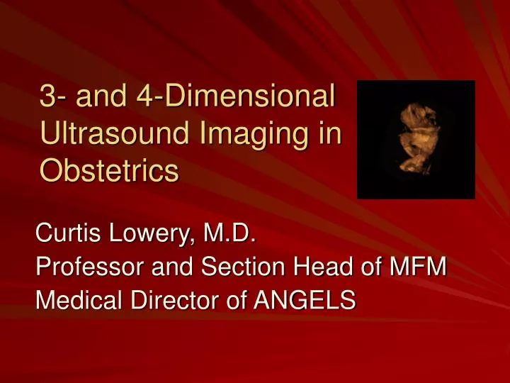
3- and 4-Dimensional Ultrasound Imaging in Obstetrics Curtis Lowery, M.D. Professor and Section Head of MFM Medical Director of ANGELS
2D to 3D • The mental process of converting 2D into 3D images is not an easy one, and is dependent on individual skills and training. • Therefore, it is not surprising that the skills involved in interpreting ultrasound images are not uniform and vary between practitioners.
Disparities in Diagnosis Ewigman BG, Crane JP, Frigoletto FD, LeFevre ML, Bain RP, McNellis D. Effect of prenatal ultrasound screening on perinatal outcome. RADIUS Study Group. N Engl J Med 1993; 329:821–827. LeFevre ML, Bain RP, Ewigman BG, Frigoletto FD, Crane JP, McNellis D. A randomized trial of prenatal ultrasonographic screening: impact on maternal management and outcome. RADIUS (Routine Antenatal Diagnostic Imaging With Ultrasound) Study Group. Am J Obstet Gynecol 1993; 169:483– 489. Grandjean H, Larroque D, Levi S. Sensitivity of routine ultrasound screening of pregnancies in the Eurofetus database. The Eurofetus Team. Ann NY Acad Sci 1998; 847:118–124. Levi S. Ultrasound in prenatal diagnosis: polemics around routine ultrasound screening for secondtrimester fetal malformations. Prenat Diagn 2002; 22:
Methods of 3-D Imaging • Freehand acquisition using a conventional 2-dimensional ultrasound (2DUS) • Freehand (2DUS) acquisition using a conventional 2DUS transducer with position sensing • Automated acquisition using dedicated mechanical volume probes • Real-time 3D imaging using 2D array transducers
There are three planes involved in the acquisition of a volume, A, B, and C. In this example of a scan with a fetus lying on its back the Planes are as follows: 1) A Plane - Transverse 2) B Plane - Longitudinal 3) C Plane - Horizontal The three planes always maintain a 90-degree relationship to each other. No matter where you move the transducer or how you rotate acquired volumes the three planes will maintain their 90-degree relationship. Thus it is important to understand that any adjustment to one plane will affect the other two planes.
Benefits of 3DUS • The ability to review volume data interactively after the patient has left the examination room • The possibility of using different planes of section for the evaluation of anatomic structures other than the original acquisition plane • The possibility of rotating the volume data set so that anatomic structures can be examined from different perspectives • The availability of a variety of rendering methods that allow examiners to visualize different characteristics of the same structure (eg, the same volume data set of the fetal back can reveal the external aspect of a meningomyelocele when rendered in the surface mode or, alternatively, the underlying bones when the volume data set is rendered in the maximum-intensity mode) Luís F. Gonçalves, MD,J Ultrasound Med 2005; 24:1599–1624
Benefits of 3DUS • Improved accuracy for volume measurements, including the possibility of measuring the volume of irregular objects • The possibility of standardizing ultrasound examinations • The ability to transmit data over networks for consultation in tertiary care centers • The potential to use offline software programs as an interactive educational tool
Fetal Face • Photography-like images • Facial movements • Good views 70% • Electronic fetal scalpel
Fetal Face • Multiplanar and rendered displays • Multiplanar: examiner to “navigate” through the volume data set simultaneously in the 3 orthogonal planes • Precise location of an anatomic structure or abnormality
Combined 2DUS and 3DUS • N=96 facial clefts • Concordance between prenatal and postnatal diagnoses was observed in 87.5% • Underestimated the severity of the clefts in 8.3% (8/96) of the cases • Overestimated in 4.1% (4/96). Rotten D, Levaillant JM. Two- and three-dimensional sonographic assessment of the fetal face, 2: analysis of cleft lip, alveolus and palate. Ultrasound Obstet Gynecol 2004; 24:402–411.
Comparison of 2DUS and 3DUS • N=31 facial clefts • Agreement between ultrasonographic diagnosis and neonatal outcomes • 87.1% (27/31) of the 3DUS examinations • 45.2% (14/31) of the 2DUS examinations • Overestimated the severity of the defects • 2DUS 41.9% (13/31) of the cases • 3DUS 9.7% (02/31) of the cases Johnson DD, Pretorius DH, Budorick NE, et al. Fetal lip and primary palate: three-dimensional versus two dimensional US. Radiology 2000; 217:236–239.
3-D Examination of Fetal Brain • Severity location and extent of anomalies • Visualization of corpus callosum • Improve visualization of cerebral blood flow • Topographic examination of fetal brain
Evaluation Of the Fetal Spine • A maximum intensity projection mode of • Rotation of volume sets • Measurement of the vertebral bodies • Level of spinal defect
Fetal Skeletal Dysplasias • 2DUS and 3DUS and 3D Helical CT 3D • Helical CT (94.1%) • 3DUS (77.1%) • 2DUS (51.4%) Ruano R, Molho M, Roume J, Ville Y. Prenatal diagnosisof fetal skeletal dysplasias by combining twodimensional and three-dimensional ultrasound and intrauterine three-dimensional helical computer tomography. Ultrasound Obstet Gynecol 2004; 24:134–140.
Congenital AnomaliesMixed Results • Xu et al: higher visualization rates for congenital anomalies • 3 DUS 78.0% [32/40] • 2 DUS 92.7% [38/41] • Scharf et al: 3DUS did not provide significant additional information (P < .05) • 2DUS (68.3% [28/41] • 3DUS 97.5% [39/41] Scharf A, Ghazwiny MF, Steinborn A, Baier P, Sohn C. Evaluation of two-dimensional versus three-dimensional ultrasound in obstetric diagnostics: a prospective study. Fetal Diagn Ther 2001; 16:333–341 Xu HX, Zhang QP, Lu MD, Xiao XT. Comparison of two-dimensional and three-dimensional sonography in evaluating fetal malformations. J Clin Ultrasound 2002; 30:515–525
Spatiotemporal Image Correlation(STIC) Inversion Mode
2 DUS vs. 3 DUS of Fetal Heart • In skilled hands little advantage • Transmission of volume datasets • Michailidis: 30 healthy fetuses • 76.0% (23/30) 4 chamber view • 83.3% (25/30) right ventricular outflow tract • 96.7% (29/30) left ventricular outflow tract • 80.0% (24/30) long axis views Michailidis GD, Simpson JM, Karidas C, Economides DL. Detailed three-dimensional fetal echocardiography facilitated by an Internet link. Ultrasound Obstet Gynecol 2001; 18:325–328.
2 DUS vs. 3 DUS of Fetal Heart • Viñals et al: 4DUS with STIC • Obstetricians with limited experience • Volume data sets • 100 fetuses examined • Visualization rates determined: • 4-chamber • left and right ventricular outflow tracts • 3-vessel view • trachea views Success rates: 81% - 100% Viñals F, Poblete P, Giuliano A. Spatio-temporal image correlation (STIC): a new tool for the prenatal screening of congenital heart defects. Ultrasound Obstet Gynecol 2003; 22:388–394.
3-DUS First Trimester of Pregnancy • Embryonic brain • Nuchal translucency • Faster scan times • High frequency transducers
Volumetry in Early Pregnancy: CorrelationWith Abnormal Pregnancy Outcome • Volumetric measurements: • Gestational sac • Yolk sac • Embryo • Fetus • Prediction of: • Spontaneous miscarriage • Aneuploidy 3-D Volumes of Limited Benefit!
Volumetry in Early Pregnancy: CorrelationWith Abnormal Pregnancy Outcome • Acharya and Morgan reported on a study of 81 patients with miscarriages • Mean gestational sac diameter/crown-rump length ratio • miscarriage, 3.3 [95% confidence interval (CI), 2.51–4.08] • normal pregnancies, 2.1 [95% CI, 1.67–2.63] • P = .008 • gestational sac volume/embryonic volume ratio • miscarriage, 3.3 [95% CI, 2.51–4.08] • normal pregnancies, 459.5 [95% CI, 81.8–837.2]; • P = .023) Acharya G, Morgan H. First-trimester, three-dimensional transvaginal ultrasound volumetry in normal pregnancies and spontaneous miscarriages. Ultrasound Obstet Gynecol 2002; 19:575–579.
Basic fetal biometric measurements Crown-rump length Biparietal diameter Head circumference Abdominal circumference Femur length Fetal anatomic survey Yolk sac, stomach, bladder renal area 4-chamber view of the heart Cord insertion Choroid plexuses, cerebral ventricles Genitalia upper and lower extremities, hands, feet, digits NTT thickness Evaluation of the uterus and placenta 32 Pregnancies meanGA of 12.3 ± 0.2 weeks Hull AD, James G, Salerno CC, Nelson T, Pretorius DH. Three-dimensional ultrasonography and assessment of the first-trimester fetus. J Ultrasound Med 2001; 20:287–293.
Fetal Anatomic and Biometric Survey byFirst-Trimester 3DUS • Complete biometric assessment • 3DUS 78.8% [126/160] • 2DUS 47.5% [76/160]; P < .001 • Nuchal translucency • 3DUS 96.9% (31/32) • 2DUS 37.5% (12/32) • Total scan times • 3DUS 14.7 ± 0.9 min. vs 2DUS 13.2 ± 0.4 min. • P< 0.05 • Transducer active time • 3DUS 02.7 ± 0.2 min. vs 2DUS 14.7 ± 0.9 min. • P < .001
Nuchal Translucency Thickness Measurements • NTT <3.0 mm • Statistically significant overestimation of values from transvaginal and transabdominal 3DUS • NTT >3.0 mm • Statistically significant underestimation of values from transvaginal and transabdominal 3DUS Worda C, Radner G, Lee A, Eppel W. Three-dimensional ultrasound for nuchal translucency thickness measurements: comparison of transabdominal and transvaginal ultrasound. J Soc Gynecol Investig 2003; 10:361–365.
3D Volumetric Measurements • Limbs to estimate: fetal weight to 5% birth weight • 3DUS 20/30 fetal weight to • 2DUS 6/30 • Lungs to predict: pulmonary hypoplasia • ? Small studies
Sonographic Tomography • 5 volume data sets • Fetal head • Face • Chest • Abdomen • Limbs • Examined by physicians not involved in acquisition • Complete studies 20/25 • Scan times reduced by half with 3DUS volume acquisitions (13.9 versus 6.6 minutes P< .001) Benacerraf BR, Shipp TD, Bromley B. How sonographic tomography will change the face of obstetric sonography: a pilot study. J Ultrasound Med 2005; 24:371– 378.
Maternal Bonding • 100 patients • Randomly assigned to 2DUS only or 2DUS + 4DUS • No difference in positive response rates • Maternal antenatal attachment scale • Quality and intensity of attachment • Global attachment score Rustico MA, Mastromatteo C, Grigio M, Maggioni C, Gregori D, Nicolini U. Two-dimensional vs. two- plus four-dimensional ultrasound in pregnancy and the effect on maternal emotional status: a randomizeds study. Ultrasound Obstet Gynecol 2005; 25:468–472.
Additional information in the diagnosis of congenital anomalies facial clefts neural tube defects skeletal malformations New resources for fetal examination Multiplanar Anatomic slicing Rendering modes Probable decrease in examination times New methods of fetal biometry Fetal organ volume measurements Volume dataset acquisition and transfer Education Conclusions