Download
1 / 10
100 likes | 229 Views
As I learned on Roots from my coleagues. “Follow the MAP!” www.rxroots.com. Middle mesial from Brasil. Márcia Valéria Boussada Vieira to. Middle Mesials : Connect the dots Trough the Line NO MM’s. On 2/26/06 A hmad T ehrani

E N D
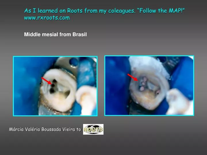
As I learned on Roots from my coleagues. “Follow the MAP!” www.rxroots.com Middle mesial from Brasil Márcia Valéria Boussada Vieira to
Middle Mesials: Connect the dots Trough the Line NO MM’s
On 2/26/06 • Ahmad Tehrani • Is there anything that alerts you to look for MM other than troughing? • Should we take in to consideration, how far apart the MB & ML canals are forinstance? • Is this anatomy discernable on the pre op x-ray? wire film? post op? • How would you recommend filing this canal so it doesn't get blocked or ledged?
On 02/27/06 • Fred Barnett • I connect the dots and trough a few mm's on every case. On the angled pre-ops, if the mesial root looks especially wide, I may be a bit more aggressive....but I always stay on the line and I am always cognizant of the furca. • When you look at Hess's work from 1928, I am humbled by the simple white lines that appear on my post-op radiographs.
Monday, February 27, 2006 2:18 PM Fred Barnett • A strategy on creating pathways to the canals is required in order to debride and disifect them. • Proper magnification and illumination to see the subtle color changes of the "Road Map" is mandatory.
“Anatomy can be wild but even the wildest can be overcome with an understanding of shape, access, and development of a thoughtful strategy that allows debridement and obturation of the clinically significant space leaving only the most minute entombed crypts remaining and unlikely to cause recurrent pathosis. “ (T. Pannkuk, 2006; ROOTS) Terry Pannkuk ‘s contribution
Monday, February 27, 2006 10:31 AM From Dr. Keneth Serota ...axial dentin has a different chromatic (colour) tone than dentin and orifices that are present within the root trunk.....that dentin is much darker and just keeping it wet magnifies the chromatic distinction.......Fred's point about troughing the line in conjunction with any combination of NaOCl, etch and a DG 16 endo explorer can assist one in finding the middle mesial canal..............access openings that truly allow light in, beveled cavo surface line angles obviate shadowing in the same way they enhance optics for photography..............each root canal system has a distinct anatomy, however, the map of the floor, the chromatic topographical outline is always in evidence regardless of the magnification or ocular apparatus.
sashi nallapati NSU endo, FL