Download
1 / 24
250 likes | 367 Views
Characterizing a -Synuclein Membrane Bound Structure. Bert Lai (Gray Group) Department of Chemistry and Chemical Engineering California Institute of Technology Thesis Defense 04/29/08. Introduction: α-Synuclein. Role of α -syn in Parkinson’s disease pathogenesis unclear

E N D
Characterizing a-Synuclein Membrane Bound Structure Bert Lai (Gray Group) Department of Chemistry and Chemical Engineering California Institute of Technology Thesis Defense 04/29/08
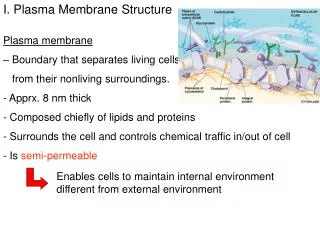
Introduction: α-Synuclein • Role of α-syn in Parkinson’s disease pathogenesis unclear • Aggregates form amyloid fibrils • major component of Lewy Bodies deposits • found in substantia nigra of Parkinson’s disease victims • Three single point mutations • (A30P, A53T, and E46K) • Early onset familial Parkinson’s • disease Substantia nigra Parkinson’s Disease Normal
α-Synuclein Sequence • Three interesting regions: • Non β-Amyloid Component (NAC) region (Residues 61-95) • Commonly found in Alzheimer’s disease aggregates • Acidic C-terminal region (Residues 96-140) • 7 imperfect repeats of 11 amino acids • Second and seventh residues of each repeat are mostly KTKEGV • Typically found in apolipoproteins Ulmer, T. S.; Bax, A.; Cole, N. B.; Nussbaum, R. L. J. Biol. Chem.2005, 280, 9595-9603.
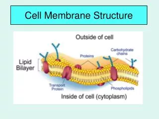
Introduction: α-Synuclein and Membrane • Synaptic vesicle-associated protein • Oligomers of α-syn involved in membrane permeabilization • Structure: • In solution: unfolded with structural preference • In SDS micelles/acidic small unilamellar vesicles (SUVs): • highly helical 20-50 nm 1 nm SDS micelles SUVs
NMR Model of α-Synuclein in Micelles Hydrophilic Tail (Gly93 – Ala140) 2nd Helix (Lys45 – Thr92) Linker (Lys38 – Thr44) 1st Helix (Val3 – Val37) • Formation of α-helices at • residue 3 to 37 (1st helix) • residue 45 to 92 (2nd helix) • Formation of linker between 2 helices • Unstructured hydrophilic tail Ulmer, T. S.; Bax, A.; Cole, N. B.; Nussbaum, R. L. J. Biol. Chem.2005, 280, 9595-9603.
Phospholipid Vesicles • EPR: Helical structure in the 7 N-terminal 11-amino acid repeats Lipid exposed Solvent exposed Jao, C. C.; Der-Sarkissian, A.; Chen, J.; Langen, R. Proc Natl. Acad. Sci.2004, 101 (22), 8331-8336.
Phospholipids The CD spectra show that acidic lipid is needed for α-syn to form helical structures. 1:1 POPC:POPA SUVs were made 1-Palmitoyl-2-Oleoyl-sn-Glycero-3-Phosphocholine (POPC) 1-Palmitoyl-2-Oleoyl-sn-Glycero-3-Phosphate (POPA)
Trp-Tyr(NO2) pairs W4-Y94 Helix #2 94 94 4 4 Helix #1 ro = 26 Å 10 < r < 40 Å
FET Kinetics & Polypeptide Conformations Lyubovitsky, J. G.; Gray, H. B.; Winkler, J. R. J. Am. Chem. Soc.2002, 124 (19), 5481-5485.
Steady State Fluorescence Studies Intensity Wavelength (nm) W4 is in a more hydrophobic environment when associated with SDS or SUV Amount of quenching by Y(NO2): SDS > SUV > Buffer
Time-resolved Fluorescence Studies 94 4 90 % NaPi 80 % P(r) SUV It 40 % SDS Distance (Å) Time (ns) The two helices are closer together in SDS micelles. 4
Summary P(r) SUV SDS Distance (Å) 94 4 • Extended helix is elucidated when α-syn is in association with SUVs • Similar to the EPR model Some extended structure remains • α-syn forms compact structures • when SDS micelles are introduced • Similar to the NMR model
C-terminal Tail • locates in residues 96-140 • is unstructured in the presence of membrane mimic • is highly negatively charged (10 Glus & 5 Asps) • modulates aggregation • binds to • metal ions, e.g. calcium, copper • microtubule-associated proteins 1B • polyamines • Aging → Oxidative Stress → Calcium Dysregulation Ulmer, T. S.; Bax, A.; Cole, N. B.; Nussbaum, R. L. J. Mol. Biol.2005, 280, 9595-9603.
Theory 1: C-terminal tail becomes β-sheet Theory 2: C-terminal tail rigidified by binding calcium ion Study C-terminal tail behavior in the presence of POPC:POPA with various Ca2+ concentration Stage 1: Trp-only mutants Stage 2: Donor-acceptor pair Tamamizu-Kato, S.; Kosaraju, M. G.; Kato, H.; Raussens, V.; Ruysschaert, J.-M.; Narayanaswami, V. Biochemistry2006, 45, 10947-. de Laureto, P. P.; Tosatto, L.; Frane, E.; Marin, O.; Uversky, V. N.; Fontana, A. Biochemistry2006, 45, 38, 11523-.
94 101 136 125 • DNP (N-Dinitrophenyl Phosphatidylethanolamine) • Br (6,7) POPC (1-Palmitoyl-2-Stearoyl(6,7-dibromo)-sn-Glycero-3-Phosphocholine) Quencher located on the end group Headgroup/hydrocarbon boundary and quencher:3.5 Å McIntosh, T. J.; Holloway, P. W. Biochemistry1987, 26, 1783-1788.
Trp Only Steady-state Fluorescence Calcium Titration Curves (in SUVs) W125 NATA [Ca2+] [Ca2+] W101 W136 [Ca2+] [Ca2+]
CD CD shows calcium ion does not affect the structure of α-synuclein. Time-resolved Fluorescent Lifetimes W101 seems to be more associated with the membrane, but not deeply inserted.
W101 → DNP Fluorescence Transfer Rate DNP + Ca2+ DNP A shift to shorter lifetimes is observed when calcium ions were added. P(r) Log(k)
Theory 1: C-terminal tail becomes β-sheet Theory 2: C-terminal tail rigidified by binding calcium ion Tamamizu-Kato, S.; Kosaraju, M. G.; Kato, H.; Raussens, V.; Ruysschaert, J.-M.; Narayanaswami, V. Biochemistry2006, 45, 10947-. de Laureto, P. P.; Tosatto, L.; Frane, E.; Marin, O.; Uversky, V. N.; Fontana, A. Biochemistry2006, 45, 38, 11523-.
80 Val Thr Gly Val Thr Ala Val Ala Gln Lys 90 Thr Val Glu Gly Ala Gly Ser Ile Ala Ala 94 100 Ala Thr Gly Phe Val Lys Lys Asp Gln Leu 101 110 Gly Lys Asn Glu Glu Gly Ala Pro Gln Glu 113 120 Gly Ile LeuGluAsp Met Pro Val AspPro 125 130 Asp Asn Glu Ala TyrGlu Met Pro Ser Glu 136 140 Glu Gly Tyr Gln AspTyrGluProGlu Ala Effective calcium binding has been shown for sequences DXXXD. Acidic Residues Mutation Sites Ca Binding
113 74 125 94 101 136 • Seven mutants were expressed: • W94/Y113 6) W101/Y125 • W94/Y125 7) W101/Y136 • W94/Y136 • W125/Y136 • W101/Y74 Graphic made in PyMol
80 Val Thr Gly Val Thr Ala Val Ala Gln Lys 90 Thr Val Glu Gly Ala Gly Ser Ile Ala Ala 94 100 Ala Thr Gly Phe Val Lys Lys Asp Gln Leu 101 110 Gly Lys Asn Glu Glu Gly Ala Pro Gln Glu 113 120 Gly Ile LeuGluAsp Met Pro Val AspPro 125 130 Asp Asn Glu Ala TyrGlu Met Pro Ser Glu 136 140 Glu Gly Tyr Gln AspTyrGluProGlu Ala Effective calcium binding has been shown for sequences DXXXD. Acidic Residues Mutation Sites Ca Binding Binding sites for Calcium in solution is found between residue 101-113 & residue 125-136