Download
1 / 132
1.41k likes | 2.14k Views
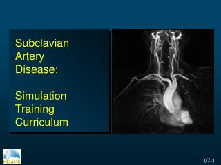
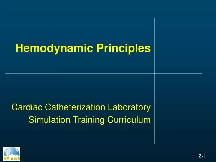
Hemodynamic Principles Cardiac Catheterization Laboratory Simulation Training Curriculum. Invasive Cardiovascular Curriculum. Hemodynamic Principles Electrocardiography Pressure waveforms Right and left heart catheterization Valvular heart disease Pericardial disease

E N D
Hemodynamic PrinciplesCardiac Catheterization LaboratorySimulation Training Curriculum
Invasive Cardiovascular Curriculum Hemodynamic Principles Electrocardiography Pressure waveforms Right and left heart catheterization Valvular heart disease Pericardial disease Hypertrophic cardiomyopathy Dampened and ventricularized waveforms
Ischemic Heart Disease Acute Inferior Myocardial Infarction Adapted from http://www.ecglibrary.com/
Ischemic Heart Disease Acute Anterior Myocardial Infarction Adapted from http://www.ecglibrary.com/
Ischemic Heart Disease Acute Posterior Myocardial Infarction Adapted from http://www.ecglibrary.com/
Ischemic Heart Disease Old Inferior Myocardial Infarction Adapted from http://www.ecglibrary.com/
Ischemic Heart Disease Acute Myocardial Infarction in the Presence of Left Bundle Branch Block Adapted from http://www.ecglibrary.com/
Hypertrophy Patterns Left Ventricular Hypertrophy (LVH) Adapted from http://www.ecglibrary.com/
Hypertrophy Patterns Mitral Stenosis Adapted from http://www.ecglibrary.com/
Hypertrophy Patterns Right Atrial Hypertrophy Adapted from http://www.ecglibrary.com/
Hypertrophy Patterns Left Ventricular Hypertrophy in the Presence of Left Anterior hemiblock Adapted from http://www.ecglibrary.com/
Atrioventricular (AV) Block First Degree AV block Adapted from http://www.ecglibrary.com/
Atrioventricular (AV) Block 2 to 1 AV Block Adapted from http://www.ecglibrary.com/
Atrioventricular (AV) Block Complete Heart Block Adapted from http://www.ecglibrary.com/
Atrioventricular (AV) Block Atrial Fibrillation and Complete Heart Block Adapted from http://www.ecglibrary.com/
Bundle Branch Block Right Bundle Branch Block Adapted from http://www.ecglibrary.com/
Bundle Branch Block Left Anterior Hemiblock Adapted from http://www.ecglibrary.com/
Bundle Branch Block Left Bundle Branch Block Adapted from http://www.ecglibrary.com/
Bundle Branch Block 'Trifasicular' Block Adapted from http://www.ecglibrary.com/
Supraventricular Rhythms Sinus Bradycardia Adapted from http://www.ecglibrary.com/
Supraventricular Rhythms Sinus Tachycardia Adapted from http://www.ecglibrary.com/
Supraventricular Rhythms Atrial Bigeminy Adapted from http://www.ecglibrary.com/
Supraventricular Rhythms Atrial Flutter Adapted from http://www.ecglibrary.com/
Supraventricular Rhythms Wolf-Parkinson-White Syndrome With Atrial Fibrillation Adapted from http://www.ecglibrary.com/
Ventricular Rhythms Ventricular Bigeminy Adapted from http://www.ecglibrary.com/
Ventricular Rhythms Polymorphous ventricular tachycardia (Torsade de pointes) Adapted from http://www.ecglibrary.com/
Ventricular Rhythms Ventricular Fibrillation Adapted from http://www.ecglibrary.com/
Invasive Cardiovascular Curriculum Hemodynamic Principles Electrocardiography Pressure waveforms Right and left heart catheterization Valvular heart disease Pericardial disease Hypertrophic cardiomyopathy Dampened and ventricularized waveforms
Pressure Waveforms Resolution of a normal ventricular pressure curve (top) into its first 10 harmonics by Fourier analysis. Hϋrthle Manometer Grossmans “Catheterization” 7th Ed. pg. 135.
Factors that Influence the Magnitude of Reflected Waves Grossmans “Catheterization” 7th Ed. pg. 135.
Wave Pressure Generator Left ventricular pressure measured with a fluid-filled standard catheter and micromanometer (catheter tip pressure manometer) in a patient undergoing cardiac catheterization Adapted from Nichols et al. Cardiovasc Res 1978; 12:566
Practical Evaluation of Dynamic Response Characteristics of a Catheter-transducer System
System for Pressure Measurement with excellent Frequency Response Strain Gauge Pressure Transducer Strain Gauge Connection of the Wheatstone Bridge
Technique for Measurement of a Patient’s Anteroposterior Diameter
Transformation of Arterial Pressure Waveform with Transmission to the Periphery in a Healthy 30-year old man
Invasive Cardiovascular Curriculum Physiologic Monitoring Electrocardiography Pressure waveforms Right and left heart catheterization Valvular heart disease Pericardial disease Hypertrophic cardiomyopathy Dampened and ventricularized waveforms
Invasive Cardiovascular Curriculum Physiologic Monitoring Electrocardiography Pressure waveforms Right and left heart catheterization Valvular heart disease Pericardial disease Hypertrophic cardiomyopathy Dampened and ventricularized waveforms
Aortic Stenosis • Most common valve lesion being considered for surgical replacement in United States • AVR improves survival of symptomatic patients (especially in patients over 65) • Symptomatic state and LV function improve or normalize in most patients after surgery
Aortic Stenosis: Etiologies Age < 70 Age 70 Passik CS, et al, Mayo Clin Proc 62:119, 1987
Aortic Stenosis: Natural History Ross J Jr, Braunwald E. Circulation 38[Suppl V]:61, 1968
Aortic Stenosis and CHF • Life expectancy ~ 2 years • Clinical improvement with AVR is not guaranteed • With diminishing ejection fraction, both perioperative and overall mortality increase
AVR and Preoperative EF Morris, et al. Ann Thorac Surg 1993: 56:22-30
AS and LVEF 35% • Operative Risk of AVR is high • 30-day mortality 9% • Worse in setting of concomitant CAD (>= 2 vessel disease associated with RR 4.6) • However, clinical outcomes are still good, in general, with surgery • 76 % show improved EF • 88% have improvement in symptoms (NYHA class) Connolly, H, et al. Circulation. 1997; 95: 2395-2400.
Pitfalls in Low-Output AS • Valve Area calculation is flow dependent: • “Pseudostenosis” is possible if Cardiac output is less than 4.5 L/min and gradient is low • Aortic Valve Resistance may be better indicator of severity (< 275 dynes/sec/cm-5) Cannon, et al. JACC 1992; 20:1517-23