Download
1 / 27
280 likes | 490 Views
NEOPLASIA Lecture 5. Maha Arafa,MD,KSFP Abdulmalik Alsheikh , M.D, FRCPC. Objectives. Define tumor grade and clinical stage. Define cachexia and its cause.

E N D
NEOPLASIALecture 5 MahaArafa,MD,KSFP AbdulmalikAlsheikh, M.D, FRCPC
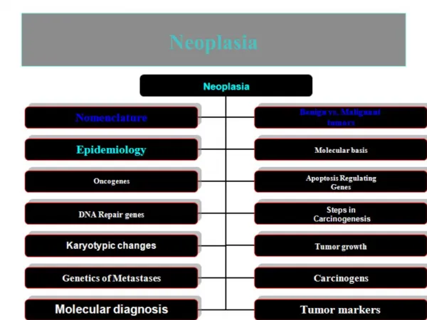
Objectives Define tumor grade and clinical stage. Define cachexia and its cause. Define paraneoplastic syndrome, and know examples of tumors associated with endocrinopathies, osseous changes, and vascular and hematologic changes. Be familiar with the general principles, value, procedures, and applications of biopsy, exfoliative and aspiration cytology, and frozen section. List some examples of tests used to diagnose cancer by immunohistochemistry and flowcytometry. Discuss the use of molecular diagnostic testing in the setting of cancer diagnosis, prognosis, minimal residual disease evaluation, and diagnosis of hereditary predisposition
Host defense • Tumor Antigens: • Tumor-specific antigens: present only on tumor cells • Tumor-associated antigens: present on tumor cells and some normal cells
Host defense • Tumor antigens may: • Result from gene mutations: P53, RAS • Be products of amplified genes: HER-2 • Viral antigens: from oncogenic viruses • Be differentiation specific: PSA in prostate • Oncofetal antigens: CEA, Alpha fetoprotein • normal embryonic antigen but absent in adults….in some tumors it will be re-expressed, e.g: colon ca, liver cancer
Host defense • Antitumor mechanisms involve: • Cytotoxic T lymphocytes • Natural killer cells • Macrophages • Humoral mechanisms : • Complement system • Antibodies
Clinical features • Tumours cause problems because : • Location and effects on adjacent structures: (1cm pituitary adenoma can compress and destroy the surrounding tissue and cause hypopituitarism). (0.5 cm leiomyoma in the wall of the renal artery may lead to renal ischemia and serious hypertension). • Tumors may cause bleeding and secondary infections • lesion ulcerates adjacent tissue and structures
EFFECT OF A TUMOR ON THE HOST • Secondary fracture
Clinical features • Effects on functional activity • hormone synthesis occurs in neoplasms arising in endocrine glands: • adenomas and carcinomas of β cells of the islets of the pancreas produce hyperinsulinism. • Some adenomas and carcinomas of the adrenal cortex elaborate corticosteroids. • aldosterone induces sodium retention, hypertension and hypokalemia • Usually such activity is associated with benign tumors more than carcinomas.
Clinical features Cancer cachexia • Usually accompanied by weakness, anorexia and anemia • Severity of cachexia, generally, is correlated with the size and extend of spread of the cancer. • The origins of cancer cachexia are multifactorial: • anorexia (reduced calorie intake) • increased basal metabolic rate and calorie expenditure remains high. • general metabolic disturbance
Clinical features Paraneoplastic syndromes • They are symptoms that occur in cancer patients and cannot be explained by spread of tumor or by elaboration of hormones indigenous to the tissue from which the tumor arose . • They are diverse and are associated with many different tumors. • They appear in 10% to 15% of pateints. • They may represent the earliest manifestation of an occult neoplasm. • They may represent significant clinical problems and may be lethal. • They may mimic metastatic disease.
Clinical features • The most common paraneoplastic syndrome are: • Hypercalcemia • Cushing syndrome • Nonbacterial thrombotic endocarditis • The most often neoplasms associated with these syndromes: • Lung and breast cancers and hematologic malignancies
Paraneoplasticsyndromes Syndrome Mechanism Example Cushing'sSyndrome ACTH-like substance Lung small (oat)cellcarcinoma Hypercalcemia Parathormone-like substance Lung squamouscell carcinoma Renal cell carcinoma Breast carcinoma Hyponatremia Inappropriate ADH secretion Lung small (oat) cellcarcinoma Polycythemia Erythropoietin-like substance Cerebellar haemangioma Renal cell carcinoma Trousseau'sSyndrome Hypercoagulable state Various carcinomas Hypoglycemia Insulin-like substance Various carcinomas and sarcomas CarcinoidSyndrome 5-hydroxy-indoleacetic acid (5-HIAA) Metastatic malignantcarcinoid tumors
Clinical Features • Grading : • Grade I, II, III, IV • Well, moderately, poorly differentiated, anaplastic • Staging : • Size • Regional lymph nodes involvement • Presence or absence of distant metastasis • TNM system
Grading of Malignant Neoplasms Grade Definition I Well differentiated II Moderately differentiated III Poorly differentiated IV Nearly anaplastic
Oat cell carcinima of the lung Undifferenciated carcinoma Grade IV Poorly differentiated neoplasms have cells that are difficult to recognize as to their cell of origine Higher grade means: a lesser degree of differentiation and the worse the biologic behavior Adenocarcinoma of the colon Well differenciated carcinoma A welldifferentiated neoplasm is composed of cells that closely resemble the cell of origin.
Clinical Staging • T (primary tumor): T1, T2, T3, T4 • N (regional lymph nodes): N0, N1, N2, N3 • M (metastasis): M0, M1
Staging of Malignant Neoplasms Stage Definition Tis In situ, non-invasive (confined to epithelium) T1 Small, minimally invasive within primary organ site T2 Larger, more invasive within the primary organ site T3 Larger and/or invasive beyond margins of primary organ site T4 Very large and/or very invasive, spread to adjacent organs N0 No lymph node involvement N1 Regional lymph node involvement N2 Extensive regional lymph node involvement N3 More distant lymph node involvement M0 No distant metastases M1 Distant metastases present
Laboratory Diagnosis • Morphologic methodes • Biochemical assays • Molecular diagnosis
Laboratory Diagnosis • Microscopic Tissue Diagnosis • the gold standard of cancer diagnosis. • Several sampling approaches are available: • Excision or biopsy • Frozen section • fine-needle aspiration • Cytologic smears
cytologic methods Slide 8.56
Laboratory Diagnosis • Biochemical assays: • Useful for measuring the levels of tumor associated enzymes, hormones, and tumor markers in serum. • Useful in determining the effectiveness of therapy and detection of recurrences after excision • Elevated levels may not be diagnostic of cancer (PSA). • Only few tumor markers are proved to be clinically useful, example CEA and α- fetoprotein.
Laboratory Diagnosis • Molecular diagnosis • Polymerase chain reaction (PCR) example: detection of BCR-ABL transcripts in chronic myeloid leukemia. • Fluorescent in situ hybridization (fish) it is useful for detecting chromosomes translocation characteristic of many tumors Both PCR and Fish can show amplification of oncogenes (HER2 and N-MYC)
Molecular diagnosis • DNA microarray analysis • Expression of thousands of • genes are studied. • Different tissue has different pattern of gene expression. • Powerful tool useful for subcategorization of disease e.g. Lymphoma • - confirmation of morphologic diagnosis • - illustration of genes involved in certain disease and possible therapy.