Download

1 / 22
220 likes | 224 Views
Does transfusion of stored red blood cells cause clinically important adverse effects? A critical question in search of an answer and a plan.

E N D
Does transfusion of stored red blood cells cause clinically important adverse effects? A critical question in search of an answer and a plan.
RBC transfusions are lifesaving, RBCs can be stored under refrigeration in preservativesolutions such as such as AdSol solution 1, 3, or 5 for up to 42 days.1 These storage solutions have met the licensure requirements mandating that RBCs transfused at the end of the approved storage period should have at least 75% of the cells remaining in the circulation 24 hours after infusion, and that the hemolysis in the stored bag be 1%.
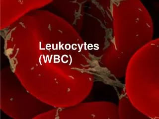
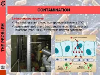
Effect of WBC contamination and leukoreduction exposure to the acidic conditions of storage and refrigeration,become activated and release cytokines, which can lead to theirdelivery at high concentrations during transfusion. In addition, during routine storage of RBCs, lipids accumulate in the plasmafraction that can prime neutrophils, causing neutrophil-mediatedcytotoxicity of human pulmonary endothelial cells, which has beenimplicated as the mechanism of lung injury in transfusion-related • acute lung injury (TRALI).
Heme and hemeoxygenase system • Spitalnik have argued that the reticuloendothelial system is burdened with an acute, large dose of RBCs after transfusion of stored RBCs with potential adverse immunological consequences. • These investigators also pointed out that mostRBC clearance of stored human units occurs within the first hour after transfusion and showed that transfusion of stored, but notfresh, mouse leukoreduced RBCs results in rapid clearance ( 2 hours) of 16% of transfused blood by themonocyte/macrophage system.
Delivering such large amounts of hemoglobin to monocytes/macrophages is likely to affect macrophage plasticity, which is viewed as a spectrum of activation states ranging from the classic proinflammatory(M1) state, which is induced by the Th1 cytokine IFN- and bacterial components such as lipopolysaccharides,andthe alternative activated (M2) state, which is associated with the resolution phase of inflammation and driven by IL-4, IL-10, TGF, and glucocorticoids.
HO-1 is considered immunosuppressive because it was shown to block the maturation of dendritic cells and to inhibit proinflammatory and allogeneicimmune responses. Dendriticcells treated with HO-1 and CO can inhibit ROS and the production of proinflammatory cytokines such as IL-12, IL-6, TNF-, and IFN type I. Conversely, the antiinflammatory IL-10 cytokine is increased after HO-1 overexpression or CO, and CO alone has been shown to inhibit T-lymphocyte proliferation. This has led to the proposal that HO-1 and CO may mediate immune tolerance by favoring the induction of master immunosuppressor cells, the so-called regulatoryTcells (Tregs). Therefore, the activation of HO-1 expression can be thought of as a critical parameter to switch the classical proinflammatoryactivity of macrophages into an immunoregulatory one.
Hod et al hypothesized that the pro-oxidant effects of iron released after acute clearance of stored RBCs may be responsible for some of the harmful effects of RBC transfusion after prolonged storage. Consistent with this hypothesis, pretreatment with the Food and Drug Administration–approved iron chelator deferoxamine before transfusion reduced cytokine production, possibly through its antioxidant activity.
Microparticles and C-mediated regulation RBC membrane loss resulting in the release of microparticles into the supernatant is considered a potential procoagulant and proinflammatory component of the storage lesion. Many complementsystem components and immunoglobulins are enriched in RBC microparticles, which is consistent with a proinflammatory potential. In addition, microparticles express the procoagulantphosphatidylserine and the number of phosphatidylserine-enriched RBC microparticles increases with storage time.
It may be that the transfusion of stored blood can create an inflammatory microenvironment due to the delivery of high concentrations of lipids and microparticles accumulated during storage.
Blood Components Preparation and Storage The standard procedure of blood products preparation from whole blood donation is as follows: once collected in plastic bags containing citrate phosphate dextrose (CPD) anticoagulant, whole blood is centrifuged in order to separate blood cells according to their size and density. Red blood cells (RBCs) settle, while plasma remains on the top. White blood cells and platelets (PLTs) form a “buffy coat” layer at the interface. Finally, the three components are distributed among the sterile inter-connected blood bags by applying a semi-automated pressure to the centrifuged bag containing the original whole blood donation.
The temperature is a particularly important storage parameter regarding the viability and the quality of products intended for transfusion. Supplemented with an additive solution, generally a saline-adenine-glucose-mannitol (SAGM) solution, RBCs can be stored for up to 42 days from +2 °C to +6 °C, in order to preserve the functionalities of erythrocytes. On the contrary, PLT are stored from +20 °C to +24 °C up to 5 days, with sufficient agitation to permit a good oxygenation and to prevent platelet aggregation.
First, some biochemical changes related to the energy metabolism occur. It appears that components such as ATP, which is necessary for multiple cellular processes, and 2,3-DPG, which plays an important role in oxygen release, rapidly decrease during the storage. ATP level is considerably diminished after 5 weeks of storage while the 2,3-DPG is almost null after 2 weeks of storage .The low concentration of 2,3-DPG increases hemoglobin affinity for oxygen, which cannot be delivered anymore. However, these levels are rapidly recovered in blood circulation after transfusion of the erythrocyte concentrate. It is also known that intracellular sodium and potassium levels are altered through storage. Indeed, Na+/K+ pumps are inactive at 4 °C, thus allowing high sodium influx and potassium loss
Then, RBCs storage induces biomechanical changes. The erythrocytes rheological properties, such as shape, deformability, aggregability, and intracellular viscosity are altered during storage .All these changes impact the red blood cells ability to pass through microvessels, thus altering their oxygenation capacities. This is very problematic in blood banking, and neither SAGM nor phosphate-adenine-glucose-guanosin-saline-mannitol (PAGGSM), the two mostly used additive solutions, are described to prevent these storage lesions
Finally, modifications also take place at the protein level. Indeed, erythrocytes are subjected to oxidative lesions, which result in protein oxidative modifications ,hemichrome formation and Band 3 clustering. Storage-induced protein degradation appears to be greatly reduced when oxygen is removed and blood is stored under helium .Antonelou et al. have shown that erythrocyte proteins are less oxidized when RBCs are stored in CPD-SAGM compared to storage in CPD-Adenine .
The normal platelet discoid shape (also referred as resting shape) is found to be lost after 5 to 7 days of storage at 22 °C. At this storage time, mainly spherical or fragmented platelets remain. Granule release and platelet activation occur during PLT storage, as indicated by the accumulation of β-thromboglobulin and platelet factor 4 in the storage medium, and the increase in surface levels of P-selectin (CD62P), respectively. Shapira et al. have also shown that platelet prothrombinase activity and membrane phosphatidylserine exposure are enhanced during PCs blood banking storage
ايمنوگلوبين ها • آنتي ژنهاي پيري در اثر مكانيسم اكسيداسيون تشكيل ميشوند