Download
1 / 13
240 likes | 2.21k Views
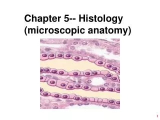
Microscopic Female Anatomy. Cross Section of Ovary. The development of follicles from their initial stage as a primordial follicle through their final stage as a Graafian follicle and ovulation. Each follicle houses one primary oocyte, which is surrounded by granulosa cells. Primordial

E N D
The development of follicles from their initial stage as a primordial follicle through their final stage as a Graafian follicle and ovulation. Each follicle houses one primary oocyte, which is surrounded by granulosa cells. Primordial Primary Secondary Tertiary Graafian Ovulation Folliculogenesis
Follicles of the Ovary • Primordial follicles • Present at birth • Decrease in number as female matures • Primary follicles • Remain at this stage until puberty • Most never ovulate and undergo atresia
Follicles of the Ovary • Secondary Follicles • Increase in granulosa cell layers • Formation of Zona Pellucida • Theca Interna and Externa • Tertiary Follicles • Maturing follicles • Antrum formation • fluid filled space • Liquor folliculi
Follicles of the Ovary • Graafian Follicle • Similar structures as tertiary follicle • Layers & fluid increase -large, blister-like structure forms on surface of ovary • Capable of ovulation • Dependent on LH surge
Luteogenesis and Luteolysis Luteogenesis: the formation of a corpus luteum Luteolysis: the regression of a corpus luteum
Luteogenesis and Luteolysis • Corpus hemorrhagicum (CH) • newly ruptured follicle • essentially a blood clot • Corpus luteum (CL) • Yellow structure • temporary endocrine gland • progesterone • Corpus albicans (CA) • remains after CL regresses
Ovarian Abnormalities Cystic follicles: the failure of a Graafian follicle to ovulate. It persists on the ovary and continues to grow. The follicle still produces high amounts of estradiol and can cause the animal to show a continued estrus behavior Luteal Cysts: occur when theca interna cells do not fill the cavity that is created upon ovulation (the rupturing of the follicle). They can cause abnormal estrous cycles and early embryonic mortality. Persistent Corpus Luteum: the failure of the CL to regress. It causes a suppression of the estrous cycle due its resemblance to pregnancy (i.e., progesterone is still produced and the body falsely identifies this state as a pregnancy).