Download
1 / 37
370 likes | 376 Views
Anatomy” and Function of Prokaryotes Dr. Hala Al- Daghistani.

E N D
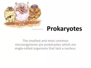
Bacteria have many sizes and several shapes. Most bacteria range from 0.2 to 2.0 um in diameter and from 2 to 8 um in length. They have a few basic shapes: 1. Spherical coccus (plural:cocci)2. Rod-shaped bacillus (plural: bacilli)3. Spiral.- Cocci that remain in pairs after dividing are called Diplococci- those that divide and remain attached in chain like patterns are called Streptococci- those that divide in two planes and remain in groups of four are known as Tetrads. - Those that divide in three planes and remain attached in cubelike groups of eight are called Sarcinae.- Those that divide in multiple planes and form grapelike dusters or broad sheets are called Staphylococci
Most bacilli appear as Single rods. Diplobacilli appear in pairs after division , and Streptobacilli occur in chains. Some bacteria are rod and look so much like cocci that they are called Coccobacilli
Spiral bacteria have one or more twists.Bacteria that look like curved rods are called Vibrios.Others, called Spirilla, have a helical shape, and fairly rigid bodies.Another group of spirals are helical and flexible; they are called Spirochetes.Unlike the spirilla, which use external appendages called flagellato move, spirochetes move by means of axial filaments, which resemble flagella but are contained within a flexible external sheath.
The shape of a bacterium is determined by heredity. Genetically, most bacteria are Monomorphic; that is, they maintain a single shape.Some bacteria, are genetically Pleomorphic. Which means they can have many shapes, not just one.
Prokaryote “Anatomy” Overview Cell envelope: Collectively all the structures outside from the plasma membrane.
Gram-negative Cell Wall Lipid A of LPS acts as endotoxin; O polysaccharides are antigens for typing, e.g., E. coli O157:H7 Gram neg. bacteria are less sensitive to medications because outer membrane acts as additional barrier. LPS layer = outer layer of outer membrane (protein rich gel-like fluid)
Gram StainDifferential staining to distinguish cell wall types.(Christian Gram 1884)
Acid-fast Cell Walls • Genus Mycobacterium and Nocardia • mycolic acid (waxy lipid) covers thin peptidoglycan layer • Do not stain well with Gram stain use acid-fast stain
Bacteria with No Cell Wall: Mycoplasmas • Instead, have cell membrane which incorporates cholesterol compounds (sterols), similar to eukaryotic cells • Cannot be detected by typical light microscopy pleomorphic Mycoplasmas
Mycoplasmascannot be detected by the naked eye or even by typical light microscopy. The morphology of mycoplasma colonies is often likened to a "fried-egg" because they form a dense central core, which penetrates downward into the agar, surrounded by a circular spreading area that is lighter in color.Mycoplasmapneumoniaeis a very small bacterium, in the class Mollicutes. This class of organisms lack a peptidoglycancell wall present on all other firmicute bacteria. Instead, it has a cell membrane which incorporates cholesterol compounds, similar to eukaryotic cells.- Lacking a cell wall, these organisms are resistant to the effects of penicillins and other beta-lactam antibiotics, which act by disrupting the bacterial cell wall.M. pneumoniae has one of the smallest genomes known, with 816 kilobase pairs (kbs).
Damage to the Cell Wall- One way the cell wall can be damaged is by exposure to the digestive enzyme lysozyme. Lysozyme is active on the major cell wall components of most gram positive bacteria, making them vulnerable to lysis. When bacteria are treated with 1) enzymes that are lytic for the cell wall e.g. lysozyme or 2) antibiotics that interfere with biosynthesis of peptidoglycan, wall-less bacteria are often produced. Usually these treatments generate non-viable organisms. Wall-less bacteria that can not replicate are referred to as 1. Spheroplasts,gram negative bacteria (when an outer membrane is present) or 2. Protoplasts, gram positive bacteira (if an outer membrane is not present).
Some members of the genus Proteus, as well as other genera, can lose their cell walls and swell into irregularly shaped cells called L forms. They may form spontaneously or develop in response to penicillin or lysozyme . L forms can live and divide repeatedly or return to the walled state.- L
Cytoplasmic Matrix and Structures • Cytoplasmic proteins • Ribosomes • Nucleoid • Inclusion Bodies • Glycogen (G) • Poly-β-hydroxybuterate (lipid) • Cyanophycin (N) granules • Carboxysomes (ribulose I.5 -diphosphatecarboxylase, CO2 fixation) • Gas vacuoles (vesicles) • Polyphosphate granules(volutin) • Magnetosomes are inclusions of iron oxide (Fe304) • Sulfur granules
Inclusion Bodies Magnetosomes = orientation Gas Vacule = buoyancy
Ribosomes • Subunits made of proteins and ribosomal ribonucleic acids (rRNA). • 30S and 50S must bind together to form a complete and functional ribosome. • The two subunits “sandwich” messenger RNA (mRNA). • As it moves along the mRNA, the genetic code is “translated” into a polypeptide by the directed polymerization of amino acids. • Transfer RNAs (tRNA) shuttle the amino acids to the ribosome as needed. • Hence, ribosomes are responsible for protein synthesis.
Plasma Membrane • Membranes are lipid bilayers (hydrophilic outside and hydrophobic inside) • Functions: • Selective permeable barrier into (out of) cytoplasm. • Transport for nutrients, excretion, secretion • Sensing the environment & signaling a response • Metabolic processes (respiration; photosynthesis …) • Excretion of hydrolytic exoenzymes • Site of biosynthesis of DNA, cell wall polymers and membrane lipids.
Structures External to the Cell Wall • S-layer:extremely well organized layer of protein or glycoprotein subunits that forms a rigid mesh, next to cell wall. Functions in • -adherence • Protect the bacteria from enzyme and change in Ph • Contribute to virulence (antiphagocytosis, anticomplements) Glycocalyx:means sugar coating; often polysaccharide or polypeptide layer external to the cell wall. • Capsules: organized, consolidated, well attached. • Slime Layer: unorganized; loose; removed easily. • Function in attachment; protection; virulence.
Structures External to the Cell Wall • Flagella: • Mostly made of flagellin. • Filament thick (20 nm) & long (10-20 µm). • Varied locations on cell: • Fimbriae: • 1000’s of thin (~5 nm) & short appendages of helical proteins. • Attachment to (specific) surfaces. • Sex Pili: • 1-10 slightly larger than fimbriae. • Only in cells with a fertility plasmid (F factor), called donors. • Attaches to like cells without F factor, called recipients. • Facilitates genetic transfer between cells; with recipient gaining the F factor and possibly other genes. peritrichous monotrichous amphitrichous lophotrichous
Endospore Formation Dormant, tough, non-reproductive structure germination vegetative cells Spore forming genera: Clostridium Resistance to UV and radiation, desiccation, lysozyme, temperature, starvation, and chemical disinfectants Relationship to disease Sporulation: Endospore formation Germination: Return to vegetative state
Endospores: • Resting stage during “lean or stressful times”. • Resistant protein coat! • Develop in different locations of vegetative cell: • Schaeffer – Fulton Stain: free; sub-terminal; central; terminal Young (24 h) Old (96 h)
Depending on the species, the endospore might be located terminally(at one end), subtermillally(near one end, or celltrallyinside the vegetative cell. When the endospore matures, the vegetative cell wall ruptures (lyses), killing the cell, and the endospore is freed.Most of the water present in the forespore cytoplasm is eliminated by the time sporulation is complete, and endospores do not carry out metabolic reactions.
The highly dehydrated endospore core contains only DNA, small amounts of RNA, ribosomes, enzymes, and a few important small molecules. The latter include a large amount of an organic acid called dipicolillic acid which is accompanied by a large number of calcium ions.Endospores can remain dormant for thousands of years. An endospore returns to its vegetative state by a process called Germination. Sporulation in bacteria is not a means of reproduction. Thisprocess does not increase the number of cells.
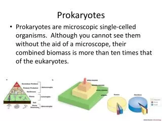
Procaryotic and Eucaryotic CellsBiologists recognize the existence of two fundamentally different types of cells in the microbial world, called Procaryotic andEucaryotic cells.1. Eucaryotic cells have a "true" nucleus (the region of the cell that contains genetic information or DNA) that enclosed in a nuclear membrane2. Procaryotic cells are said to have a "primitive" nucleus because their DNA is not enclosed within a nuclear membrane. The nuclear region of a procaryotic cell is sometimes referred to as a nucleoid, but never as a nucleus. 3. Eucaryotic cells are always bounded by a membrane, just as prokaryotic cells are.4. Some eucaryotic cells are also surrounded by a cell wall, but eucaryotic cells do not have capsules. Mitochondria are present in nearly all eucaryotic cells and produce the cell's energy by breaking down food. Chloroplasts, in contrast, are present only in plants and algae and are used in photosynthesis, the process through which the organism uses energy from the sun to build sugars.
Cytoplasm • Composed largely of water, together with proteins, nucleic acid, lipids and small amount of sugars and salts • Ribosomes: numerous, 15-20nm in diameter with 70S; distributed throughout the cytoplasm; sensitive to streptomycin and erythromycin site of protein synthesis • Plasmids:extrachromosomal genetic elements • Inclusions: sources of stored energy, e,g volutin
Plasmid Plasmids are small,circular/line,extrachromosomal,double-stranded DNA molecules。They are capable of self-replication and contain genes that confer some properties,such as antibiotic resistance,virulence factors。Plasmids are not essential for cellular survival. Inclusions of Bacteria • Inclusions are aggregates of various compounds that are normally involved in storing energy reserves or building blocks for the cell. Inclusions accumilate when a cell is grown in the presence of excess nutrients and they are often observed under laboratory conditions. granulose