Download
1 / 37
370 likes | 603 Views
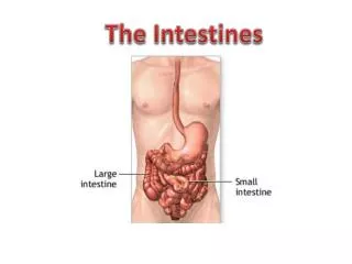
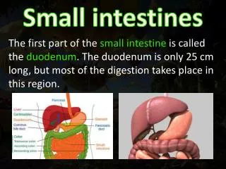
The Intestines. Small and large intestines. Some disease processes are common to both In other ways they are functionally and pathologically different Small bowel – villous surface specialised for food absorption Large bowel – water and electrolyte absorption. Intestinal immune system.

E N D
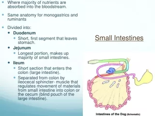
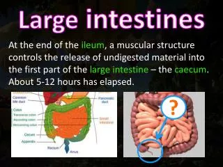
Small and large intestines • Some disease processes are common to both • In other ways they are functionally and pathologically different • Small bowel – villous surface specialised for food absorption • Large bowel – water and electrolyte absorption
Intestinal immune system • Large amounts of lymphoid tissue throughout intestines • Specialised MALT. Circulating cells of this system “home” to gut • B-cells specialised for Ig A production • T-cells include intraepithelial lymphocytes
Congenital abnormalities • Atresia or stenosis (e.g. imperforate anus) • Meckel diverticulum – terminal ileum. Can contain gastric/pancreatic mucosa leading to ulceration/perforation
Hirschprung’s disease • Developmental disorder characterised by lack of ganglion cells in nerve plexus of gut leading to loss of motility • Aganglionic segment extends proximally from rectum for a variable distance • Important cause of childhood constipation
Diarrhoea • Hard to define • Some mechanisms • Secretory – stimulated by toxins (e.g. cholera) • Exudative – more severe mucosal damage with bloody stool (e.g. typhoid) • Malabsorption – bulky fatty stools
Infective causes of diarrhoea • 12,000 deaths per day in developing countries (mainly children) • Viruses • Bacteria • Parasites
Viral enteritis • Rotavirus – cytopathic effect on mature enterocytes, replaced by immature cells with loss of absorptive function (infants mainly) • Adenovirus Cause a degree of villous flattening and increased intraepithelial lymphocytes
Bacterial enteritis/enterocolitis • Mechanisms: • Toxin – either formed by proliferating bacteria in gut or ingested directly with food • Enterotoxins – disturb metabolic function of epithelium (cholera) • Cytotoxins – kill epithelial cells (Shigella) • Adherence to and invasion of gut tissue (Shigella, E.coli)
Salmonella enteritis • Many Salmonella species ( e.g. enteritidis) exist in animal (poultry) reservoirs and cause diarrhoea through poorly cooked food • S. typhi is confined to humans so spread is purely faecal-oral
Pathogenesis of Salmonella diarrhoea • Organisms invade epithelial cells and macrophages • Typhoid in particular associated with systemic disease (fever, rash, pain, prostration and GI haemorrhage) • Septicaemia preceeds recolonisation of gut and gallbladder • Reabsorbed through Peyer’s patches which ulcerate (effect of immune reaction)
Pathology of typhoid • Longitudinal ulcers • Perforation • Haemorrhage • Cholecystitis • Multiorgan disease – liver, kidney, bone, striated muscle
Carriers • Infection can linger in bone and particularly gallbladder • “Typhoid Mary”
Cholera • Vibrio cholerae • Noninvasive • Produces enterotoxin which stimulates enterocyte secretion of salt and water • Morphological changes not prominent, some villous stunting
Shigella, Campylobacter • Invasive • Acute enteritis/colitis with dysentery • Acute inflammatory cell infiltration of mucosa with crypt abscesses
E.coli • Very common (travellers diarrhoea) • Very variable pathogenesis • Enterotoxigenic subtypes (E0157 associated with haemolytic uraemic syndrome) • Enteroinvasive subtypes (Shigella – like)
Other bacteria • Clostridia – C.difficile causes antibiotic associated colitis (pseudomembranous) • Yersinia – mesenteric adenitis and ileo-colic ulceration
Intestinal tuberculosis • Primary – ingestion of organism in unsensitised host. Can cause severe ulcero-inflammatory disease with perforation • Secondary – swallowing of infected sputum • Most common in terminal ileum and jejunum • Complications – obstruction, fistula.
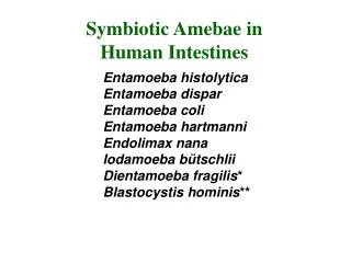
Protozoal enterocolitis • Giardia – very common worldwide • Coccidia • Cryptosporidiosis • Isospora These organisms associate with cell membrane. Water borne. Very common with HIV
Amoebiasis • Simple tissue invading unicellular organism • Deep flask-shaped ulcers
Amoebic dysentery • Organisms can be seen in inflammatory exudate • Can spread by blood stream giving an amoebic liver abscess
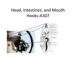
Nematodes • Ascaris – can physically obstruct intestine. Also liver abscess, pneumonia • Hookworms – mucosal attachment causes erosion and bleeding • Strongyloides – invade wall of gut and can persist for life causing life-threatening systemic disease later (HIV)
Schistosomiasis • S. mansoni (rarely S. haematobium) • Mainly affects the colorectum • Larva migrate to liver and mature before moving to submucosal vessels of gut where eggs are laid • Proctitis, oedema, haemorrhage
Schistosomiasis • Ova detectable in rectal biopsy • Chronic inflammation with eosinophils • Can lead to scarring/obstruction
HIV associated disease • Diarrhoea is a big problem • Opportunistic infection (candida, cryptosporidia, cytomegalovirus, Mycobacterium avium-intracellulare, strongyloides, leishmaniasis) • HIV itself causes enteropathy • Kaposi’s sarcoma
HIV • Multiple pathologies common
Malabsorption • Defective absorption of fats, proteins, carbohydrates and other nutrients (vitamins, minerals) • Clinical hallmarks are diarrhoea (sometimes very fatty – steatorrhoea), malnutrition
Malabsorption • Normal process involves • Intraluminal digestion • Terminal digestion (disaccharidases and peptidases on epithelial brush border) • Trans-epithelial transport
Causes of malabsorption (1) • Defective intraluminal digestion • Pancreatic insufficiency (e.g. chronic pancreatitis) • Loss of bile flow (biliary obstruction) • Nutrient preabsorption by bacterial overgrowth (e.g. in surgical “blind loops”)
Causes of malabsorption (2) • Loss or abnormality of epithelial surface • Tropical sprue • Chronic infective conditions (e.g. TB) • Extensive surgical resection of small bowel • (Other chronic inflammatory conditions – Crohn’s disease, coeliac disease)
Causes of malabsorption (3) • Lymphatic obstruction • TB • Lymphoma
Causes of malabsorption (4) • Infection • Acute enteritis of any kind • Parasites • Tropical sprue
Effects on small bowel • Atrophy of villi • Inflammation • Increased intraepithelial lymphocytes • Means different things in different populations
Inflamed atrophic small bowel • Europe – coeliac disease • Africa tropical sprue • Bacterial overgrowth following enteritis • Can be treated with antibiotics
Crohns disease Involves any part of GI tract Abnormal areas are interspersed with normal “skip lesions” Ulcerative colitis Confined to colon Inflammation continuous from rectum Idiopathic inflammatory bowel disease
Microscopy • Crohn’s inflammation is transmural, sometimes granulomatous • Ulcerative colitis inflammation is mucosal
Inflammatory bowel disease • A major problem in Europe/N. America • Apparently uncommon in Africa but may be masked by the predominance of infective disease

![STOMACH, INTESTINES, RECTUM [SURGICOSE]](https://cdn4.slideserve.com/8061806/medical-instruments-medical-instruments-dt.jpg)