Download
1 / 20
220 likes | 362 Views
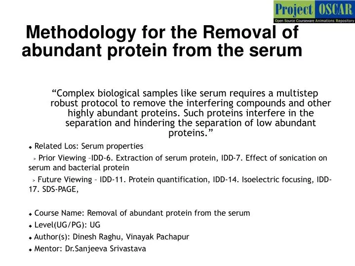
Methodology for the Removal of abundant protein from the serum.

E N D
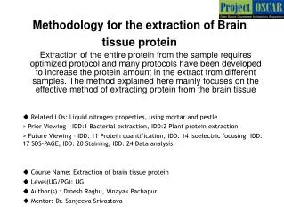
Methodology for the Removal of abundant protein from the serum “Complex biological samples like serum requires a multistep robust protocol to remove the interfering compounds and other highly abundant proteins. Such proteins interfere in the separation and hindering the separation of low abundant proteins.” • Related Los: Serum properties > Prior Viewing –IDD-6. Extraction of serum protein, IDD-7. Effect of sonication on serum and bacterial protein > Future Viewing – IDD-11. Protein quantification, IDD-14. Isoelectric focusing, IDD-17. SDS-PAGE, • Course Name: Removal of abundant protein from the serum • Level(UG/PG): UG • Author(s):Dinesh Raghu, Vinayak Pachapur • Mentor: Dr.Sanjeeva Srivastava
Learning objectives 1 • After interacting with this Learning Object, the learner will be able to: • Define the techniques and the materials involved in the serum depletion • Distinguish the abundant proteins in the serum • Interpret the result before and after the serum depletion. • Infer the steps involved to perform the experiment. • Assess the troubleshooting steps involved in the experiments. 2 3 4 5
Master Layout 1 High abundant proteins in the serum (Side:5) 2 Depletion Column (Side:6-8) 3 Addition of Binding buffer (Side:9-10) Removal of High abundance proteins (Side:11-13) 4 Sample storage (Side:14) 5
Definitions and Keywords 1 Serum : Serum is the yellowish part of the blood, devoid of cells and clotting factors. Serum contains all blood proteins and secreted proteins which can be used for the study. Depletion column:The affinity column that are used to trap the high abundance proteins like IgG, albumin etc in serum are called depletion column. Depleted serum:Serum devoid of high abundance proteins. Albumin:It constitute 60% of the blood protein and it is produced by the liver. It acts as a carrier of hydrophobic compounds like bile, fatty acid etc. Haptoglobin:The protein that bind to free hemoglobin and reduces it oxidative property. Immunoglobin G:IgG is mostly present in the serum as a result immune response in the system and prevent the infection of any infectious agent 2 3 4 5
Audio Narration Description of the action Step 1: T1:High abundant proteins in the serum 1 2 3 The animator should show a tube containing brownish liquid labeled as serum. Animate in such a way that the user should click on the liquid to get the composition of it. Draw a red ring and label as IgG, Orange circle as alpha-1 antitrypsin, green as IgA, blue as Transferrin, Yellow-Albumin and brown -haptoglobin . Animate similarly as shown The serum consists of high abundance proteins such as IgG,alpha-1 antitrypsin, IgA, transferrin, haptoglobin which will interfere during 2D separation. These proteins mask the movement of low abundant proteins 4 5
Description of the action Audio Narration Step 2: T2: Depletion Column 1 2 3 The animator should draw a freezer and user opening the freezer to take box labeled as depletion column. Animate in such a way so that the user should click on the box to open and take a depletion column wrapped. Instruct user to remove the wrap and animate the action of removing the wrap to take out column. Remove the depletion column from the freezer in advance to the pre-treatment. 4 5
Step 2: T2: Depletion Column 1 2 3 4 Centrifuge rotor 5
Description of the action Audio Narration Step 2: T2: Depletion Column 1 The animator should draw a centrifuge as shown in the figure. Animate in such a way that user clicks on lid to open it and keep the column inside the rotor (with lots of holes) as shown. The animator should animate like the user should click on setting and set 800 rpm , 5minutes and click “enter” and animate like closing the lid and click “start”. Show a clock running for 5 minutes. Once the 5 minutes is done the user should open the lid by clicking “open” and remove the column out. Please include the buttons like enter, set, start, open in the centrifuge. Centrifuge the column for 5 minutes at 800 rpm to remove storage buffer. 2 3 4 5
Step 3: T3: Addition of Binding buffer 1 2 3 4 pipette 5
Description of the action Audio Narration Step 3: T3: Addition of Binding buffer 1 Add binding buffer to the column and centrifuge. Buffer treatment is done to activate the bonding property of resins inside the column The animator should draw a freezer and a hand opening the freezer and take box labeled as Binding buffer. Animate in such a way so that the user should click on the box to open and take a binding buffer wrapped. Instruct user to remove the wrap and animate the action of removing the wrap to take out binding buffer. The animator should draw a pipette as shown in the previous slide and set 400ul in the pipette to draw binding buffer to transfer it onto the column. Now let user close the column and keep it in centrifugation and setting as shown in slide:10. 2 3 4 5
Description of the action/ interactivity Audio Narration (if any) Instruct user to take out column from centrifuge, zoom the column to show some liquid level at the bottom of the tube, animate to discard the liquid into waste labeled bottle. Instruct user to add 50ul of serum to the column, when user clicks on the pipette, animate the pipette action and later place the tube on ice for 5min. Please redraw the figure Step 4: T4:Removal of High abundance proteins 1 2 3 Discard the liquid collected in the column below, now the column is ready to carry out depletion. Add the serum to the binding column and keep in ice for 5mins. 4 5
Description of the action/ interactivity Audio Narration (if any) Animate flow of serum into the column, show protein like IgG, albumin binding to the column with no further movement and the small abundance protein having a free flow and getting eluted at faster rate. The animator should animate like the crescent shaped proteins binding to the pink rings in the column and blue square shaped and yellow triangle shaped coming out of the column . Draw as given in the previous slide. Step 4: T4:Removal of High abundance proteins 1 2 3 High abundance protein get attracted and bind to the affinity column while other low abandance proteins does not bind and get eluted at faster rate. This step helps in depleting the sample. 4 5
Audio Narration Description of the action Step 4: T4:Removal of High abundance proteins 1 2 3 The animator should draw a centrifuge as shown in the figure. Animate in such a way that user clicks on lid to open it and keep the column inside the rotor (with lots of holes) as shown. The animator should animate for user to click on setting and set 800 rpm , 30 sec 4 degree Celsius temperature and click “enter” and animate like closing the lid and click “start”. Show a clock running for 30 sec. Once the 30 sec is done the user should open the lid by clicking “open” and remove the column out. Please include the buttons like enter, set, start, open in the centrifuge. Centrifuge the column for 30 sec at 800 rpm. Centrifuge helps to collect the unbound protein sample at the bottom of the column. 4 5
Description of the action Audio Narration Step 5: T5: Sample Storage 1 2 3 Animate in such a way that the user should remove the column from the centrifuge and take the liquid from the bottom of the tube. The animation should be like the user draw the serum from the pipette and transfer that to the new tube. And keeping it at -20 C freezer in a box Transfer the serum to the new tube and store it till further usage. For more information please go through the future viewing IDD from slide:1. 4 5
Button 01 Button 02 Button 03 Slide 14 Slide 5 Slide 6-8 Slide 9-10 Slide 11-13 Introduction Tab 01 Tab 02 Tab 03 Tab 04 Tab 05 Tab 07 Name of the section/stage Animation area Interaction 1: • Let user run the sample without removing abundant proteins. • Instruction: animate to display the final gel image with more proteins all compressed together. 1 image without depletion and second one is depleted serum Interactivity area Instructions/ Working area Credits
Questionnaire: APPENDIX 1 Question 1: Albumin constitute-------------------- percent of the blood a)65% b)70% c)60% d)45% Answer:c)60% Question 2: Haptoglobin bind to a)Hemoglobin in erythrocytes b)Hemoglobin in liver cells c)Free hemoglobin d)Oxygen Answer:c)Free hemoglobin
Questionnaire: APPENDIX 1 Question 3: Most abundant of the immunoglobin is a)IgM b)IgG c)IgA d)IgG and IgM Answer: IgG Question 4: Depletion column works by a)Affinity binding b)Hydrophobic interaction c)Ion exchange d)protein-protein exchange Answer:a)Affinity binding
Questionnaire: APPENDIX 1 Abundant proteins in the serum includes • Albumin • Haptogloblin • IgG • All the above Answer: All the above
APPENDIX 3 Summary Depletion of high abundance proteins from the serum is the most important step that improves the quality of the separation of the serum proteome and enable proper separation of the low abundance proteins. Such step has to be done as per suggested protocol to ensure better result. • Albumin and IgG represent greater than 60-80% of the total protein in human serum content. Therefore, removal of albumin and IgG from serum samples, prior to electrophoresis, improves the resolution of lower-abundance proteins in two ways: by enabling visualization of proteins that co-migrate with albumin and IgG; and by removal of a large portion of the total serum protein, which allows an increase in the protein load of the low-abundant proteins.
APPENDIX 2 Links for further reading Reference websites: 2DE Tutorials by Angelika Görg : http://www.wzw.tum.de/blm/deg/ Books: Biochemistry by Stryer et al., 5th edition Biochemistry by A.L.Lehninger et al., 3rd edition Biochemistry by Voet & Voet, 3rd edition GE Healthcare handbook 2D electrophoresis principles and method, by Angelika Görg, 3rd edition Research papers: Chen JH, Chang YW, Yao CW et al. Plasma proteome of severe acute respiratory syndrome analyzed by two-dimensional gel electrophoresis and mass spectrometry.Proc Natl Acad Sci U S A2004, 7;101(49):17039-44. Eymann C, Dreisbach A, Albrecht D. A comprehensive proteome map of growing Bacillus subtilis cells. Maldonado AM, Echevarría-Zomeño S, Jean-Baptiste S. et al. Evaluation of three different protocols of protein extraction for Arabidopsis thaliana leaf proteome analysis by two-dimensional electrophoresis. Proteomics 2008, 71(4):461-72.