Download
1 / 1
10 likes | 98 Views
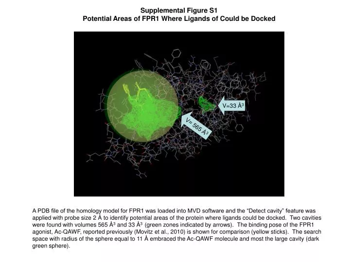
V=33 Å 3. V= 565 Å 3. Supplemental Figure S1 Potential Areas of FPR1 Where Ligands of Could be Docked.

E N D
V=33 Å3 V= 565 Å3 Supplemental Figure S1 Potential Areas of FPR1 Where Ligands of Could be Docked A PDB file of the homology model for FPR1 was loaded into MVD software and the “Detect cavity” feature was applied with probe size 2 Å to identify potential areas of the protein where ligands could be docked. Two cavities were found with volumes 565 Å3 and 33 Å3 (green zones indicated by arrows). The binding pose of the FPR1 agonist,Ac-QAWF, reported previously (Movitz et al., 2010) is shown for comparison (yellow sticks). The search space with radius of the sphere equal to 11 Å embraced the Ac-QAWF molecule and most the large cavity (dark green sphere).