Download
1 / 8
80 likes | 215 Views
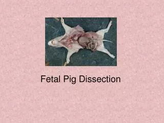
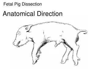
Fetal Pig Dissection Image Sets. *All Structures must be CLEARLY identified by pins or by power point arrows placed in the images. (Group grade given-All members get the same grade). External Features to ID (Unit 1 ) Image Set #1. Nares Eyes & Nicitating membrane Tongue Pinnae Thorax

E N D
Fetal Pig Dissection Image Sets *All Structures must be CLEARLYidentified by pins or by power point arrows placed in the images. (Group grade given-All members get the same grade)
External Features to ID (Unit 1)Image Set #1 • Nares • Eyes & Nicitating membrane • Tongue • Pinnae • Thorax • Trunk • Umbilical cord • Teats • Urogenital opening • Scrotum (male) or Genital papilla (female)-Pg. 9 • Anus • Measure length to estimate age of pig (use string and measure from the tip of the nose to the base of the tail) ****Use table on pg. 7 to estimate age.
Muscle ID (Three lists)Image Set #2 I. Lateral View Neck, Shoulder, and Upper Arm(See pg. 20) Triceps Deltoids Brachioradialis Trapezius Extensor digitorum lateralis Masseter Extensor digitorum communis Extensor carpi ulnaris II. List of Muscles – Ventral View (Pg. 21) Sternohyoid Pectoralis Major Brachiocephalic Pectoralis minor Latissimus Dorsi Serratus ventralis III. List of Muscles – Lateral View (See Pg. 22) Biceps Femoris External Oblique Gluteus maximus Semitendinosis Gluteus medius (medialis)
Exploration of the NeckImage Set #3 • Sternohyoid Muscle • Larynx • Thyroid gland • Thymus gland • Trachea • Jugular vein • Carotid artery
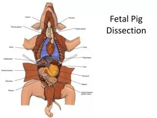
Exploration of the Thoracic Cavity Image Set #4 • Lungs (7 lobes)-Label Left & Right Side • Diaphragm • Heart • Pericardium • Apex • Coronary artery & vein • Aortic arch • Atrium • Ventricles • Pulmonary vessels (arteries & veins) • Septum
Image Set #5 - Structures of the Digestive System • Hard palate • Soft palate • Epiglottis • Esophagus • Liver(be able to name the 4 lobes) • Stomach(be able to identify cardiac region & pyloric region) • Spleen • Gall Bladder • Mesenteric Arteries • Pancreas • Small intestine (Duodenum, Jejunum, & Ileum) • Large intestine segments (A, T, D, Sigmoid) • Cecum/Spiral colon (Fig.22 on p.35) *Isolate the small & Large intestines *Measure the length of the digestive tract (Stomach to the anus)
Image Set #6 Further Exploration of Circulatory System • Locate & Identify the following Vessels: • Carotid Arteries, Jugular Veins (already done in image se #3) • Subclavianartery (branches off the aortic arch) • Brachial artery (upper extremities) • Abdominal Aorta & Vena Cava (inferior) • Iliac artery • Femoral artery (lower Extremities) • Mesenteric Arteries & Veins • Portal Vein **Know where these blood vessels can be found: -Gastric -Tibial -Hepatic -Radial -Pulmonary -Caudal
Urinary & Reproductive Systems (Image Set #7) • Kidney (cross section of left kidney) • Renal cortex (outer layer) • Renal medulla & • Renal pelvis (inner layer) • Adrenal gland • Ureter • Urinary Bladder • Renal artery & renal vein • Female (uterine horns & ovary) • Male (vas deferens & testes) ***Review the diagrams for the brain (pgs. 50 & 51)