Download
1 / 46
620 likes | 1.13k Views
Cytogenetics: Karyotypes and Chromosome Aberrations . Chapter 6. Chromosome Number. The number and appearance of chromosomes is an important characteristic in genetic analyses. Table 6-1, p. 121. Chromosome Shape.

E N D
Cytogenetics: Karyotypes and Chromosome Aberrations Chapter 6
Chromosome Number • The number and appearance of chromosomes is an important characteristic in genetic analyses. Table 6-1, p. 121
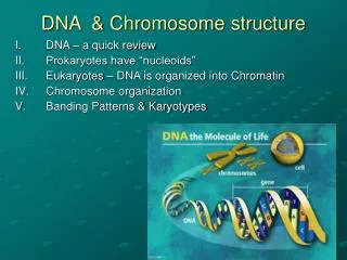
Chromosome Shape • As chromosomes condense and become visible during cell division, certain structural features can be recognized • Centromere • A region of a chromosome to which microtubule fibers attach during cell division • The location of a centromere gives a chromosome its characteristic shape
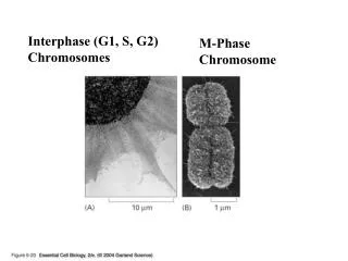
Human Chromosomes • Replicated chromosomes at metaphase consist of sister chromatids joined by a single centromere Fig. 6-1, p. 122
Centromere Location • Metacentric • A chromosome that has a centrally placed centromere • Submetacentric • A chromosome whose centromere is placed closer to one end than the other • Acrocentric • A chromosome whose centromere is placed very close to, but not at, one end
Metaphase Chromosomes • Chromosomes are identified by size, centromere location, banding pattern
A Set of Human Chromosomes • Human chromosomes are analyzed by construction of karyotypes • Karyotype • A complete set of chromosomes from a cell that has been photographed during cell division and arranged in a standard sequence
A Human Karyotype Fig. 6-3, p. 122
System of Naming Chromosome Bands • Allows any region to be identified by a descriptive address (chromosome number, arm, region, and band)
Add a few drops of blood. Add phytohemagglutinin to stimulate mitosis. Draw 10 to 20 ml of blood. Incubate at 37°C for 2 to 3 days. Transfer to tube containing fixative. Transfer cells to tube. Add Colcemid to culture for 1 to 2 hours to stop mitosis in metaphase. Centrifuge to concentrate cells. Add low-salt solution to eliminate red blood cells and swell lymphocytes. Drop cells onto microscope slide. Digitized chromosome images processed to make karyotype. Examine with microscope. Stain slide with Giemsa. Fig. 6-6, p. 124
Metaphase Chromosomes (a) Arranged Into a Karyotype (b) Fig. 6-7, p. 125
6.3 Constructing and Analyzing Karyotypes • Different stains and dyes produce banding patterns specific to each chromosome • Karyotypes reveal variations in chromosomal structure and number • 1959: Discovery that Down syndrome is caused by an extra copy of chromosome 21 • Chromosome banding and other techniques can identify small changes in chromosomal structure
Information Obtained from a Karyotype • Number of chromosomes • Sex chromosome content • Presence or absence of individual chromosomes • Nature and extent of large structural abnormalities
Four Common Chromosome Staining Procedures This is extra info that will not be on the exam.**
Chromosome Painting • New techniques using fluorescent dyes generate unique patterns for each chromosome Fig. 6-9a, p. 127
Chromosomal Aberrations and Specific Syndromes Table 6-2, p. 126
Obtaining Cells for Chromosome Studies • Any nucleus can be used to make karyotype • Lymphocytes, skin cells, cells from biopsies, tumor cells • Sampling cells before birth • Amniocentesis • Chorionic villus sampling (CVS)
Amniocentesis • A method of sampling the fluid surrounding the developing fetus by inserting a hollow needle and withdrawing suspended fetal cells and fluid • Used in diagnosing fetal genetic and developmental disorders • Usually performed in the sixteenth week of pregnancy
Removal of about 20 ml of amniotic fluid containing suspended cells that were sloughed off from the fetus A few biochemical analyses with some of the amniotic fluid Centrifugation Quick determination of fetal sex and analysis of purified DNA Fetal cells Growth for several days in culture medium Biochemical analysis for the presence of alleles that cause many different metabolic disorders Karyotype analysis (a) Fig. 6-10a, p. 127
Chorionic Villus Sampling (CVS) • A method of sampling fetal chorionic cells by inserting a catheter through the vagina or abdominal wall into the uterus • Used in diagnosing biochemical and cytogenetic defects in the embryo • Usually performed in the eighth or ninth week of pregnancy
Chorionic villi Developing placenta Ultrasound to monitor procedure Developing fetus Bladder Uterus Chorion Catheter Amniotic cavity Rectum (a) Fig. 6-11a, p. 128
Exploring Genetics: Noninvasive Prenatal Diagnosis • Methods are being investigated to isolate fetal cells that can pass into the mother’s bloodstream (placental cells, white blood cells, immature red blood cells) for genetic testing p. 129
6.4 Variations in Chromosome Number • Changes in chromosome number or chromosome structure can cause genetic disorders • Two major types of chromosomal changes can be detected in a karyotype • A change in chromosomal number • A change in chromosomal arrangement
Changes in Chromosome Number • Polyploidy • Duplication of an entire set of chromosomes • 3N = triploid • 4N = tetraploid • Aneuploidy • Refers to a single chromosome • Trisomy = one extra chromosome (three copies) • Monosomy = missing one chromosome of a pair
A Triploid Karyotype Fig. 6-12, p. 130
A Triploid Infant Fig. 6-13, p. 131
Causes of Aneuploidy • Nondisjunction • The failure of homologous chromosomes to separate properly during meiosis • About half of all conceptions are aneuploid.
Nondisjunction Extra chromosome (n + 1) Normal division Missing chromosome (n− 1) Normal (n) Normal (n) Meiosis I Meiosis II Gametes (b) Fig. 6-14b, p. 132
Extra chromosome (n + 1) Nondisjunction Extra chromosome (n + 1) Missing chromosome (n − 1) Missing chromosome (n − 1) Meiosis I Meiosis II Gametes (a) Fig. 6-14a, p. 132
Effects of Monosomy and Trisomy • Autosomal monosomy is a lethal condition • Eliminated early in development (spontaneous abortion) • Some autosomal trisomies are relatively common • Most result in spontaneous abortion • Three types can result in live births (13, 18, 21)
Trisomy 13: Patau Syndrome (47,+13) • A lethal condition • 1 in 10,000 births Fig. 6-16, p. 133
Trisomy 18: Edwards Syndrome (47,+18) • A lethal condition • 1 in 11,000 births • 80% are females Fig. 6-17b, p. 133
Trisomy 21: Down Syndrome (47, +21) • Trisomy 21 is the only autosomal trisomy that allows survival into adulthood
6.5 What Are the Risks for Autosomal Trisomy? • The causes of autosomaltrisomy are unknown • Factors that have been proposed include: • Genetic predisposition • Exposure to radiation • Viral infection • Abnormal hormone levels • Maternal age is the leading risk factor for trisomy • 94% of nondisjunctions occur in the mother • Meiosis is not completed until ovulation • Embryo-uterine interactions that normally abort abnormal embryos become less effective
6.6 Aneuploidy of the Sex Chromosomes • More common than autosomal aneuploidy • Can involve both X and Y chromosomes • A balance is needed for normal development • At least one copy of the X chromosome is required for development • Increasing numbers of X or Y chromosomes causes progressively greater disturbances in phenotype and behavior
Turner Syndrome (45,X) • Monosomy of the X chromosome that results in female sterility. Other phenotypic characteristics but otherwise normal. • Fig 6.20 Fig. 6-20, p. 136
Klinefelter Syndrome (47, XXY) • Individuals (males) have some fertility problems but few additional symptoms • Fig 6.22 Fig. 6-22, p. 137
XYY Syndrome (47,XYY) • Affected individuals are usually taller than normal and some, but not all, have personality disorders Fig. 6-23, p. 138
6.7 Structural Alterations Within Chromosomes • Changes in the structure of chromosomes • Deletion—loss of DNA • Duplication—extra DNA • Translocation—DNA that changes location • Inversion—order of DNA changes
Structural Changes in Chromosomes Fig. 6-24, p. 139
6.9 Other Forms of Chromosome Changes • Uniparental disomy • A condition in which both copies of a chromosome are inherited from a single parent • Copy number variation • A situation in which a particular gene or chromosomal region is present in multiple copies • Fragile sites • Appear as gaps or breaks in chromosome-specific locations
Human Diseases Associated with Copy Number Variants Table 6-5, p. 143
Fragile Sites • Appear as gaps or breaks in chromosomes • One fragile site on the X chromosome is associated with a common form of mental retardation in males know as Fragile X Syndrome • Females can also have this, but the phenotypes are much more mild