Download
1 / 171
1.77k likes | 2k Views
Thoracic Surgery Back to Basic. FM Shamji 2008. Esophagus. Adult - hollow muscular tube 25 cm long 1 cm in neck, 19 - 22 cm in the mediastinum, 3 cm in the abdomen Specialized sphincters at each end LES – physiological sphincter, prevents gastroesophageal reflux

E N D
Thoracic SurgeryBack to Basic FM Shamji 2008
Esophagus • Adult - hollow muscular tube 25 cm long • 1 cm in neck, 19 - 22 cm in the mediastinum, 3 cm in the abdomen • Specialized sphincters at each end • LES – physiological sphincter, prevents gastroesophageal reflux • UES – anatomical sphincter, prevents air entry during breathing and regurgitation from esophagus into pharynx
Esophagus • Three symptoms • Heart burn • Dysphagia • Chest pain • Three operations • Antireflux repair • Esophagectomy • Esophageal myotomy
Dysphagia • Difficulty in swallowing is a WORRYING symptom; precise symptom that must never be ignored • Clinical history is most important • Progressive dysphagia with progressive weight loss are suggestive of malignancy • Intermittent dysphagia implies motility disturbance • Investigations must be thorough and quick • First comes diagnosis, then comes treatment
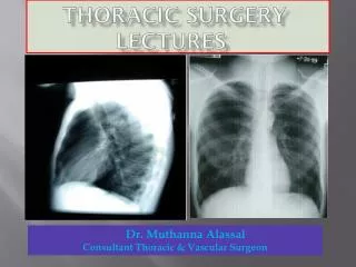
Investigations for Dysphagia • Focused clinical history, present and past • Physical examination is of secondary importance – begin with mouth, examine neck for supraclavicular lymph nodes, goitre, abdomen for hepatomegaly, ascites • CXR • Barium swallow and UGI study • Upper GI endoscopy and mucosal biopsy • Esophageal function assessment – manometry study if motility disorder is suspected • CT scan chest and abdomen
Implications of Delayed Diagnosis • Failure to thrive and weight loss • Recurrent aspiration pneumonia • Bronchiectasis • Lung abscess • Lung fibrosis • Empyema • Retention esophagitis • Local extension of cancer • Aortic-esophageal fistula • Tracheal-esophageal fistula
Dysphagia – Case 1 • A barium swallow was performed on this elderly 80-year-old man who had difficulty in swallowing. • What is the diagnosis? • What are the two important reasons for treatment? • What treatments are possible?
Zenker’s Diverticulum • Pharyngoesophageal Diverticulum in the Neck • Dysphagia and Recurrent Pulmonary Aspiration • Treatment is always Surgical • Cricopharyngeal Myotomy • Myotomy alone (if small) • Myotomy plus diverticulectomy (if large) • Acquired condition – 80% of patients are >50 years age • Pulsion diverticulum • Dysfunction of the UES – cricopharyngeus muscle • Other symptoms are: regurgitation of undigested food, choking and foul breath
Dysphagia – Case 2 • The barium study shown was obtained in a 55 year old man with a 10 year history of mild vague postprandial “indigestion” and heartburn. Within the past 6 weeks he has developed dysphagia for solid food and 10 lb weight loss. What is the next step in his management?
Mid-Esophageal Stricture • Cancer until proven otherwise • Assessment of patient • Complete history and physical examination • Heart, Lung, Liver and kidney function • Assessment of stricture • Esophagoscopy and mucosal biopsy • Rigid and flexible bronchoscopy • CT scan chest and abdomen • PET scan • Esophageal U/S • Ass
Causes of Esophageal Cancer • Adenocarcinoma • Barrett’s epithelium complicating chronic gastroesophageal reflux disorder • Squamous cell carcinoma • Excess alcohol and tobacco • Nitrosamines in pickled vegetables and cured meats • Silica in wheat • Fungus • Caustic ingestion • Achalasia • Chronic iron deficiency • Tylosis palmaris et plantaris • Remote radiation therapy to the mediastinum
Gastro-Esophageal Reflux Disorder • Backward flow of gastric juice into the esophagus • Physiological and Pathological • Lower esophageal sphincter is incompetent • Causes • Most common is idiopathic • Normal LES pressure 15 to 30 mmHg • <6 mmHg pathognomic reflux disorder • <10 mmHg reflux often present • After treatment for Achalasia • Pneumatic dilatation or distal esophagomyotomy • Scleroderma • Esophagogastrectomy • Truncal vagotomy and pyloroplasty • Prolonged Naso-gastric tube insertion
Symptoms of Gastroesophageal Reflux Disorder • Typical symptoms • Intermittent, substernal discomfort or burning sensation • Within 1 hour of eating, with exercise or recumbent • Postural regurgitation is most consistent symptom • Water brash • Atypical symptoms • Cough, laryngitis, wheezing, pulmonary due to aspiration • Dental caries due to loss of enamel
Complications of Pathological GERD • Esophagus • Reflux esophagitis • Chronic blood loss – iron deficiency anemia • Peptic stricture • Epiphrenic diverticulum • Barrett’s epithelium – risk for adenocarcinoma • True Barrett’s ulcer – perforate into mediastinum, thoracic aorta, pericardium, and airway, penetrate, bleed massively • Proximal airway • Laryngitis • Asthma • Lung • Recurrent aspiration pneumonia – fibrosis, bronchiectasis, lung abscess, empyema • Mouth – loss of tooth enamel
Esophagus • Which of the following tests is the most sensitive for the detection of pathologic gastroesophageal reflux? a. acid perfusion (Bernstein) test b. barium swallow c. 24 hr pH monitoring d. esophagoscopy e. manometric testing
discussion • 24 hour pH monitoring is now the gold standard and the most precise measurement of acid reflux. Normal pH range 4.1 and 7.0 Reflux episode is present when pH < 4.0 • Endoscopic examination to assess acid-related esophageal mucosal damage – esophagitis, stricture, columnar metaplasia • Barium swallow is for anatomical examination of esophagus, stomach and duodenum – demonstrate diverticula, hiatus hernia, esophageal stricture – a road map for safer endoscopic examination • Manometric testing of esophagus is done for suspected motility disorder as a cause of dysphagia and chest pain • Acid perfusion test will only confirm that esophagus is sensitive to acid – an attempt to reproduce symptoms
Investigations for GERD • CXR • Fixed hiatus hernia • Pulmonary parenchymal changes from recurrent aspiration • Barium study • Hiatus hernia • Stricture • Zenker’s diverticulum • Flexible esophagogastroscopy and mucosal biopsy • Hiatus hernia • Esophagitis • Columnar lined esophagus – look for Barrett’s epithelium from biopsy • Esophageal function assessment • Manometry • Impedance-pH study • 24 hr pH monitoring
Complication of GERD • A 45-year-old man presents with a history of recurrent heartburn with occasional central chest pain, and frequent episodes of postural regurgitation of gastric juice into his mouth. UGI endoscopy finding is shown. • What is the diagnosis? • Was it necessary to take mucosal biopsy? • How will the diagnosis influence future treatment?
Columnar Lined Esophagus • Acquired condition due to chronic gastroesophageal reflux • Barrett’s Epithelium – intestinal metaplasia recognized by presence of goblet cells • Malignant transformation to adenocarcinoma • Surveillance is important once dysplasia is present • High grade dysplasia is serious; within 2 years adenocarcinoma develops
Esophagus • A 40-year-old man has progressive dysphagia and 20 lb weight loss for 6 months. He had taken antacids regularly for many years for heartburn and indigestion. Esophagoscopy performed for assessment is shown. • Can you describe the findings? • Are any other tests necessary for assessment of this lesion? • What is the best treatment?
Adenocarcinoma of Esophagus • Columnar lined esophagus and distal malignant stricture • Esophageal mucosal biopsy, CT scan chest and abdomen or PET scan, esophageal ultrasound, complete blood count and serum biochemistry • Esophagectomy and reconstruction if patient is operable and cancer resectable
Esophageal Cancer • Incidence of adenocarcinoma is rising – an explosion • Gastroesophageal reflux related – not necessarily acid reflux • Develops in metaplastic Barrett’s epithelium or heterotopic gastric mucosa in esophagus • Always do mucosal biopsy of gastric cardia and distal esophagus – establish diagnosis of reflux and columnar transformation
Esophagus • A 50-year-old woman complains of nocturnal regurgitation and cough, a sour taste in her mouth, postural regurgitation, and heart burn for 5 years. She wants to consider an operation now. Which of the following is/are helpful in deciding whether to perform antireflux surgery? • failure of medical therapy to control symptoms • incompetent lower esophageal sphincter • presence of reflux related esophageal mucosal injury • increased esophageal exposure to gastric juice. • all of the above
Emergency in Esophagus • A 67-year-old man has had intermittent dysphagia, burning epigastric pain and retrosternal chest pain after meals for 2 years. For the past 24 hours, he has been unable to tolerate anything by mouth. CXR was done in the ER. • What is the diagnosis? • Does this condition constitute an emergency? • Can you describe treatment that is most appropriate?
Emergency in Esophagus • 82-year-old woman • Retrosternal chest pain, vomiting and retching for 48 hours • Similar episodes in the past 4 years
Emergency in Hiatus Hernia • Risk of Volvulus and Strangulation in incarcerated Type III or IV hernia • Early operation is mandatory to preserve stomach and prevent death from ischemic gastric necrosis and perforation • Emergency Room • Insert naso-gastric tube to decompress of stomach • Intravenous fluid resuscitation with Ringer’s Lactate • UGI Endoscopy to evaluate gastric mucosa • Stomach non-viable ---- urgent gastrectomy • Stomach viable ---- semi-urgent hiatus hernia repair and concomitant antireflux repair
Type I Type II Type III
Treatment for Hiatal Herniation • Type I • Most common 85 to 90% • Operate for severe reflux disorder refractory to optimal medical therapy for minimum of 6 months • Type II • Pure type II is rare < 1%. Operate to prevent mortality from strangulation and ischemic perforation • Type III • Mixed Type I and II • More common than Type II 6% • Needs operation for symptoms of reflux and incarceration • Type IV • Least common. Entire stomach in the chest with volvulus • Needs operation
Management of GERD • Medical Therapy is for life • Diet must be modified eating small meals and avoid late night meals • Antacids • Combination of histamine receptor blocker PM dose and proton pump inhibitor AM dose • Postural therapy by elevating head of bed 15 cm • Surgical Therapy is for failure of medical therapy • Anti reflux fundoplication
Thoracic Surgical Emergency • A 45 year old man, after a large meal and alcohol, arrives in the ER complaining of severe retrosternal and left pleuritic chest pain for 6 hours after a bout of vomiting and retching. Respiration 28, Pulse 130, BP 90/60, T 38.5 • The most likely cause of his illness is • Aortic dissection • Acute pericarditis • Myocardial infarction • Pulmonary embolism • Esophageal perforation
Esophageal Perforation • Very serious; high mortality is directly related to delay in treatment • Acute septic mediastinitis and sepsis syndrome • Iatrogenic is more common than post-emetic perforation • Operation within 1 hour - resuscitation, drainage of sepsis in the mediastinum and pleural space, adequate repair of perforation, broad spectrum antibiotic for both aerobes and anaerobes, and nutritional support
Dysphagia • This barium swallow was performed on a 33-year-old woman complaining of difficulty in swallowing both solids and liquids for 4 years with gradual worsening over time and 10 lbs weight loss. Regurgitation of undigested food has occurred. • What is demonstrated in this x-ray? • How is this diagnosis confirmed? • What is the treatment?
Esophageal Motility Disorder • Achalasia with Epiphrenic Diverticulum • Esophagoscopy is necessary to assess gastroesophageal junction – rule out peptic stricture, leiomyoma, cancer • Esophageal manometry confirms diagnosis • Preferred treatment - Distal esophagomyotomy and partial fundoplication • Pneumatic dilatation carries a risk of esophageal perforation of 4%
Achalasia • Achalasia is Greek for “without relaxation” in which the lower esophageal sphincter fails to relax before the oncoming bolus of food • Failure of the sphincter to open is due to lack of the necessary stimulus of the peristaltic wave • Motility disorder, neurogenic in origin, related to changes in the ganglion cells of the Auerbach’s plexus – degeneration, impaired function, query autoimmune in origin • Chronic condition in which treatment is for palliation of obstruction to swallowing
Definitive Diagnosis Achalasia: Manometric Features
Complications of Achalasia • Esophageal • Progressive dilatation • Retention esophagitis • Perforation • Epiphrenic diverticulum • Malnutrition • Squamous cell cancer pretreatment and adenocarcinoma post-treatment due to reflux • Trachea • Obstruction from compression due to mega-esophagus • Pulmonary • Recurrent aspiration pneumonia, lung abscess, bronchiectasis, fibrosis • Social • Withdrawn, eats alone
Mediastinum • This specimen was removed from anterior-superior mediastinum in a 45-year-old woman complaining of fluctuating weakness in arms and legs, slurring of speech, chewing difficulty, drooping of eyelids. • What is it? • How could it have been predicted? • Why was it removed? • What condition does the patient have?
Thymus Gland • Thymus gland with a thymoma • CT scan chest • Presence of tumour and for treatment of muscle weakness • Myasthenia Gravis
Myasthenia Gravis • Autoimmune disorder • Auto-antibodies against acetylcholine receptors • Reduced number of receptors destruction>synthesis • Relationship to Thymus gland • Atrophic 10% • Thymoma 8% to 15% • Hyperplasia 70% to 80%
Mediastinum • The syndrome most commonly associated with a thymoma is • Myasthenia gravis • Red blood cell aplasia • Aplastic anemia • Hypogammaglobulinemia • Cushing’s syndrome
Myasthenia gravis 30-45% Favourable prognostic factor Hypogammaglobulinemia 12% Red cell aplasia 5% Autoimmune hemolytic anemia Myositis dystrophica Myocarditis Dilated cardiomyopathy Mucocutaneous candidiasis Non-thymic malignancies Parathymic Syndromes