Download
1 / 24
240 likes | 470 Views
Microbial diseases of skin and eyes. Lesson Objectives. List the protective features of the skin and mucous membranes . Discuss the methods that pathogens use to invade the skin and mucous membranes . State locations and examples of normal flora .

E N D
Lesson Objectives • List the protective features of the skin and mucous membranes. • Discuss the methods that pathogens use to invade the skin and mucous membranes. • State locations and examples of normal flora. • Discuss selected bacterial infections of the skin and eyes; the mode of transmission and clinical symptoms of each. • List the mode of transmission, and clinical symptoms of herpesvirus and papillomavirus. • List the cutaneous fungal infections. • Discuss microbial diseases of the eye and list the microbes that cause them.
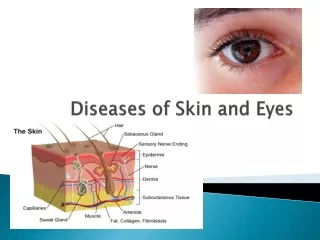
Skin—the First Line of Defense (remember???) The important stuff: 1) epidermis:stratum corneum(horned layer). Layer of dead cells that are continually sloughed off. This removes microbes.Sebaceous glands: produce oil (sebum). Sebum has a low pH (acid) which kills many pathogens. Sebum is a food source for some microbes, but it produces toxic by-products (toxic to pathogens, harmless to us!
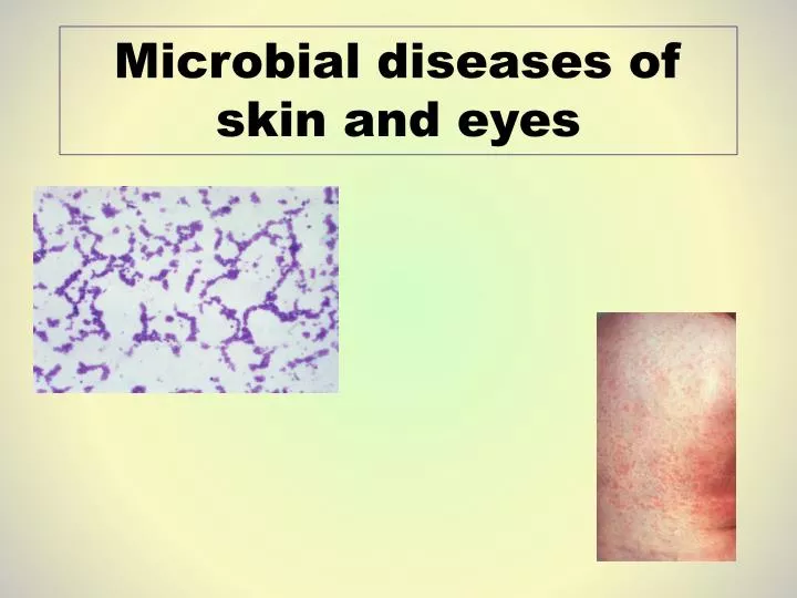
Perspiration—high salt, contains lysozyme (kills bacteria). “normal” microbes of the skin: what do we expect to find? Staphylococcus species: Gram positive cocci in clusters. Streptococcus species, especially S. pyogenes.Dermatophytes: various molds (fungi) of different species.
Although some of these species are pathogens, most of the time, they don’t cause disease (why?)
What parts of the skin would we expect to find the most microbes? • Hands___________________________________ • Face____________________________________ • Axillae__________________________________ • Anus___________________________________ • Genitalia________________________________ • Feet____________________________________
Culture of skin or tissue • Collected on a culturette, or from skin scrapings. If pus is generated, this is collected with a syringe. • Usually a Gram stain is performed. Plated on blood agar, sometimes other agar if other pathogens are suspected.
Diseases of skin—by pathogen Staphylococcus aureus: • Gram positive cocci in clusters. • Gold colonies on most media. • Beta hemolytic on blood agar • Catalase positive (H2O2 fizzes—true of all staph) Coagulase positive (produces an exotoxin that makes mammalian plasma clot.)
Virulence of Staphylococcus aureus • Exotoxins: many contribute to S. aureus’sability to cause disease:coagulase: described aboveDNase: digests cellular DNAlipase: digests fats, allows staph to colonize skin.Hemolysins (many): These are enzymes that lyse blood cells.Hyaluronidase: digests the material that holds cells in tissues together.
Staph. aureusis a notorious pathogen because… • High tolerance to environmental stress (salt tolerant, heat tolerant, facultative anaerobe. • Produces a large variety of toxins • Generally is present as normal skin bacteria • When S. aureusbreeches the First Line of Defense, it can cause many types of disease (boils, impetigo, pneumonia, food poisoning, septicemia, etc.) • Although it is usually sensitive to penicillin and related antibiotics, resistance to antibiotics is possible. MRSA.
How S. aureus invades tissue: invasiveness • Pathogen enters skin through a pore, sebaceous gland, a cut, or other portal. Fimbriae enable attachment. • Pathogen excretes exotoxins that kill or exclude host cells. The dead cell products may be a food source for the pathogen. • The pathogen reproduces, and continues to produce exotoxins that kill/exclude host cells.
Diseases of skin/tissue related to S. aureus invasive infection: • Impetigo: p. 438. Flaking “crusty” skin. • Cellulitis: p. 440. This usually involves the tissues. Sometimes occurs post-surgery. Usually uncomplicated if caught early. • Boils: infected hair follicles. Usually fluid filled skin eruptions, cause mild-severe pain. These can develop into an abscess (extend to subcutaneous tissue, and invasive).
Next pathogen: Streptococcus pyogenes (Group A streptococcus) • Gram positive cocci in chains (~ 20 cocci per chain) • Beta hemolytic on blood agar (complete hemolysis). Smaller colonies than S. aureus. • Catalase negative. Sensitive to bacitracin:
Many of the toxins produced by S. pyogenes are similar to those produced by S. aureus. Both organisms can be invasive.Necrotizing fasciitis (“flesh eating bacteria”). A very rapid invasive infection. Any invasive infection can lead to • Septicemia (bacteria in the blood. Life threatening situation) • Osteomyelitis (bacteria in the bone marrow).
Scarlet Fever • Caused by S. pyogenes that contains a bacterial virus. • Erythrogenic toxin: causes skin to become very red and rough (“sandpaper feel”) • Usually the portal of entry for scarlet fever is the respiratory tract.
Other bacteria that can enter through the skin and cause invasive infection: • Clostridium perfringens obligate anaerobe, endospore former. Causes gangrene. • Bacillus anthracis obligate aerobe, endospore former. Causes cutaneous anthrax (p. 447) • Propionibacter acnes causes acne, an infection of the sebaceous glands. Much more mild than boils.
Treatment of skin and tissue infections • Antibiotic therapy: S. aureus must always be tested for antibiotic sensitivity. • Debridement: surgical removal of necrotic and/or infected tissue. • Extreme cases (necrotizing fasciitis): amputation of affected parts.
Viral infections of the skin • Maculopapular infections (red rashes) pp. 434-438 • These are usually red, blotchy, sometimes raised, but not open. • Portal of entry is usually respiratory system. Other symptoms include fever, sore throat, and flu-like symptoms
All maculopapillary infections (except scarlet fever) are viral. They are self-limiting (they “go away”) and generally confer natural active immunity.- Measles- Rubella (a danger to pregnant women for fetus)- “fifth” disease- Coxsackie Virus- Scarlet fever
Herpesviruses and papilloma virus • Chicken pox: portal of entry: respiratory system • Begins as macular rash, develops into fluid filled vesicles. • End of infection, vesicles (pox) which open and crust over. Infection is generally self-limiting. However… • Herpesvirus (Varicella Zoster) that causes chicken pox can also cause shingles later in life.
Papilloma virus: warts Portal of entry: skin. Can spread from one part of the body to another through direct contact (auto-inoculation). Raised irregular growth.Plantar warts: on soles of the feet. Can be painful during walking.Genital warts: may appear on vulva, penis, or anus. Sexually transmitted, may be painful. In some cases, the human papilloma virus can lead to cervical or penile cancers. Shingles: infection of Varicella Zoster virus on nerve tissue. P. 443.
Fungal infections of the skin Tinea: caused by a variety of molds called dermatophytes. Usually localized, can cause itching, redness (“ringworm.” This is not a worm—it is a fungus!) Because dermatophytes do not penetrate past the epidermis, diagnosis is done through observation of symptoms. Treatments are usually topical.
Infections of the eye • Conjunctivitis (“pink eye”): infection of the conjunctiva of the eye. Heavy purulent (pus) discharge, very painful. Caused by a variety of bacteria (mostly respiratory) and viruses, primarily Hemophilisinfluenzae. • Treated with antibiotics. In Delaware, children are allowed in school/day care after 24 hours of antibiotic treatment for conjunctivitis.
Keratitis: infection of the cornea. Portal of entry: traumatic event (scratched cornea, sometimes contact lenses) Can be caused by many bacteria (S. aureus, S. pyogenes, Pseudomonas aeruginosa) and can become invasive. In this case, diagnosis and treatment must be immediate to prevent permanent damage to/loss of the eye.