Download
1 / 40
400 likes | 593 Views
Blood Laboratory Procedures. Hematology. Defined: The study of blood. Why is hematology important?. Evaluation of disease states Screening for well animals as a baseline Pre-anesthetic screening Drug-level monitoring. Hematopoiesis. Defined : The production of blood cells and platelets

E N D
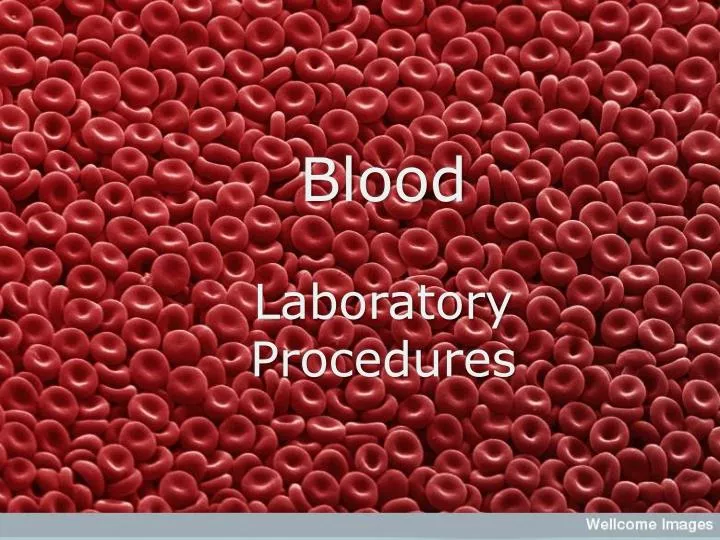
Blood Laboratory Procedures
Hematology • Defined: The study of blood
Why is hematology important? • Evaluation of disease states • Screening for well animals as a baseline • Pre-anesthetic screening • Drug-level monitoring
Hematopoiesis • Defined: The production of blood cells and platelets • All blood cells are formed from the same pluripotent stem cell!
Blood Composition • Blood is composed of fluid and cells • Fluid portion is ~90% water
Red Blood Cells:aka Erythrocytes • Erythropoiesis is the process of maturation of a RBC • Formed by the stem cell through action of the cytokine called erythropoietin (EPO) • Maturation of a RBC takes about 5 days.
Erythrocyte Life Spans • Dog ~110 days • Cat ~70 days • Cow ~ 160 days • Horse ~ 145 days • Man ~ 120 days • Mouse ~ 30 days
RBC’s continued • No nucleus • Morphology varies among species
Classification of RBC’s • RBC’s are classified the following criteria: • Cell arrangement on blood film • Size • Color • Shape • Presence of structures on erythrocytes
Classification of RBC’sCell Arrangement on Blood Film • Normal erythrocytes should lie in a nice, even, single layer on the outer-most edge of a blood film
Rouleaux Formation • Is a grouping of erythrocytes in stacks. • It can be a sign of increased fibrinogen or globulin concentration • Can be an artifact seen in blood that is held too long before preparation of blood slide or if refrigerated. • If a drop of saline is added to blood, rouleaux will disperse
AgglutinationOr Auto-agglutination • May appear as rouleaux (stacks) or in clusters • Occurs in immune-mediated disorders • An antibody coats the cell causing bridging or clumping. • If a drop of saline is added to blood, agglutination will not disperse
Classification of Blood Cells • Erythrocytes that are normally colored are called Normochromic • Polychromasia: is a variation in color • Polychromasia can appear as Hypochromasia or Hyperchromasia. • Erythrocytes that are of normal, consistent shape are called Normocytic • Anisocytosis: is a variation in size • Microcytic cells are smaller than normal cells • Macrocytic cells are larger than normal cells • Poikilocytosis: is a variation is shape • This is an “umbrella” term that is used to describe abnormally shaped cells and cannot suggest a diagnosis
Blood Collection/Handling • There are many tubes in which to collect blood • FIRST, you must know what test is being run! • Samples must be drawn before any treatment is administered. • Certain drugs can affect results • Hydration status can affect results
Where do we get the Blood? • Venous blood is almost always preferred • Jugular vein is most appropriate (and usually easiest w/ fastest flow) • Use LARGEST needle that patient can handle • Use syringe that best matches volume of sample needed.
The Vacutainer • Is composed of a needle, needle holder and collection tubes. • Use the correct size tube to minimize damage to the sample and to prevent collapse of the vein. • Fill tube to correct volume based on strength of vacuum pressure to ensure appropriate ratio of blood to anticoagulant. • ADVANTAGE: multiple samples can be collected directly into tubes without removing needle from patient.
Sample Volume • The amount of blood collected from an animal depends on the amount of serum or plasma needed as well as the hydration status of the animal. • If the animal is dehydrated, the fluid portion of the blood will DECREASE. • Example: a PCV of 50% will give a sample that is 50% cells and 50% fluid. This means that a 10mL sample will yield 5mL of fluid. A PCV of 70% will yield 70% cells and only 30% fluid so a 10mL sample will only give 3mL of fluid.
Special Note: • Enough blood should be taken to run the required tests three times. This should be enough to compensate for technician error, instrument error or the need for diluted samples.
Whole Blood • Is placed into a container with an anticoagulant added to prevent clotting. • As soon as you obtain your sample, mix the blood with the anticoagulant by using a gentle rocking motion. • Vigorously shaking your sample can cause hemolysis, otherwise known as cell destruction.
Serum or Plasma? • Serum or plasma are the fluid portion of whole blood. • Fluid portion of blood is ~90% water, 10% dissolved constituents like proteins, vitamins, carbs, hormones, etc… • Plasma DOES contain clotting factors. The clotting factors are known as fibrinogen. • Serum is plasma that has had the clotting factors removed.
Anticoagulants • Defined: Are chemicals that prevent or delay the clotting process. • Choice of anticoagulant depends on tests needed. • Sample must be well mixed before use.
Anticoagulants cont’d • Samples not tested within one hour of collection should be refrigerated. (Bring sample back to room temperature and re-mix before analysis. • Whole blood should NEVER be frozen as the freezing/thawing process can lyse the blood cells.
Red Topped Tube • Red Topped Tube: Contains no anti-coagulant. Routinely used for serum or clotted whole blood. Used for serum samples.
Serum Separator/Tiger Topped Tube • Tiger Topped (Striped) Tube/Serum Separator: • Contains no anti-coagulant. Has a yellowish “plug” of clot activation gel that separates serum from plasma when spun. Used for serum samples. (Not used for therapeutic drug level monitoring.)
Lavender/Purple Topped Tubes • Lavender Topped Tube: Contains the anticoagulant EDTA or Ethlenediamine tetraacetic acid. • Used for whole blood samples or plasma samples. • Used for complete blood counts because it does not alter cellular morphology. HOWEVER, an excess of anticoagulant in a sample may cause cells to shrink and invalidate cell counts done on automated analyzers.
Grey Topped Tube: • Contains the anticoagulant Sodium Fluoride. • Best for glucose preservation. • Interferes with many other tests performed on serum.
Blue Topped Tube • Contains the anticoagulant Sodium Citrate. • Commonly used in transfusions. • Na Citrate interferes with Na assays and many common serum tests.
Green Topped Tube • Contains the anticoagulant Heparin. • Can be used for most tests that require plasma samples. • Should never be used for differential blood film analysis because the anticoagulant interferes with the staining of the WBC’s.