Download
1 / 1
10 likes | 139 Views
#123: CD45-autoMACS depletion strategy: efficient enrichment of circulating tumor cells from peripheral blood samples of patients with various urologic cancers Meye A 1 , Bilkenroth U 2 , Schmidt U 1 , Füssel S 1 , Robel K 1 , Melchior AM 1 , Taubert H 2 , and Wirth MP 1

E N D
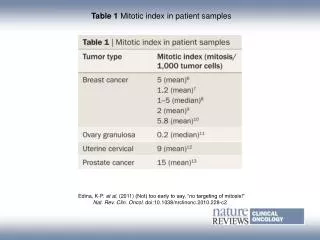
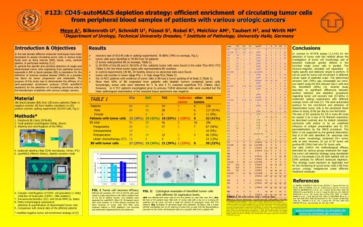
#123: CD45-autoMACS depletion strategy: efficient enrichment of circulating tumor cells from peripheral blood samples of patients with various urologic cancers Meye A1, Bilkenroth U2, Schmidt U1, Füssel S1, Robel K1, Melchior AM1, Taubert H2, and Wirth MP1 1Department of Urology, Technical University Dresden, 2 Institute of Pathology, University Halle, Germany Introduction & Objectives In the last decade different molecular techniques have been developed to assess circulating tumor cells in various body fluids such as bone marrow (BM), blood, urine, seminal plasma, or peritoneal washing [1,2]. In general the specific and sensitive detection of single and disseminated tumor cells originating from epithelial tumors especially in BM or blood samples (BS) is mandatory for the definition of minimal residual disease (MRD) as a possible risk factor for tumor progression and metastasis.The purpose of this study was to demonstrate the efficacy of a semi-automated enrichment protocol (CD45 autoMACS depletion) for the detection of circulating carcinoma cells in the bloodstream of patients with various urologic cancers. • Results • recovery rate of DU145 cells in spiking experiments: 70-88% (79% on average, Fig.1) • tumor cells were identified in 59 BS from 52 patients (5 tumor cells/positive BS on average, Table 2). • in 29-39% of the BS and in 38-53% of the patients tumor cells were found in the order PCa<RCC<TCC (Table 1) for the three tumor types with representative BS numbers • in control experiments (30 BS, 20 healthy donors) no abnormal cells were found. • tumor cell number in lower stage PCa < in high stage PCa (Table 2) • the 10 RCC patients with evidence of tumor cells in BS had a tumor grading of at least 2 (Table 2). • More than 50% of BS originated from patients with bladder tumors contained tumor cells. Surprisingly, tumor cells were detected for 5 Ta and 5 T1 resected superficial TCC (Tables 1&2). However, in 4 TCC patients investigated prior to primary TUR-B abnormal cells were counted but the histo- pathological examination of the resected tissue specimens was negative. Conclusions In contrast to RT-PCR assays [1,2,4-6] for the detection of tumor cells this method allows the investigation of tumor cell morphology and of potential molecular genetic defects in the enriched single tumor cells in parallel. The immuno-magnetic activated cell separation is highly specific and relatively simple to perform. It can be used for tumor cell enrichment in different cancer types of epithelial origin. The determined recovery rate (79%) was comparable our previ-ous report using the RCC-derived Caki-1 cells and the VarioMACS (84%) [3]. Another study describes no significant differences between positive selection and depletion procedures regarding tumor cell recovery rate (57-65%) in systematic spiking experiments with different urologic tumor cell lines [7].The semi-automated protocol for the enrichment and detection of disseminated tumor cells in the peripheral blood allows to study 20 BS per day by one technician. The lack of ICC positivity for CK in many BS could be caused i) by a loss of CK filament expression as described routinely also for distant metastatic carcinoma cells and/or ii) by an undefined influence of antigen presentation and iii) ICC permeabilization by the MACS procedure. The latter is not supported by the general observation that 8 of BS with identified CK- abnormal cells with tumor morphology contained CK+ tumor cells as well. However, 25 out of 87 tumor cell positive BS (29%) had only CK- tumor cells. Our data confirm the methodological efficacy described by various groups employed the nega-tive tumor cell selection strategy using dynabeads [7,8] or microbeads [3,9,10] both labeled with an CD45 antibody for efficient leukocyte depletion. This strategy could represent an applicable tool for the monitoring of occult tumor cells in BS from various urologic malignancies under different treatment schedules. • Material • 186 blood samples (BS) from 128 tumor patients (Table 1) • negative controls: BS from healthy volunteers (n=20) • positive controls: spiking experiments (DU145 cells, Fig.1) • Methods* • Peripheral BS (16mL EDTA-BS) • Ficoll-gradient centrifugation (650g, 20min) • Washing and centrifugation of the MNCs • leukocyte labeling (40µl CD45 microbeads; 15min, 4°C) • autoMACS (Miltenyi Biotec), deplete sensitive mode • Cytospin centrifugation of CD45- cell population (1 slide) collection of leukocytes (CD45+, DNA isolation) • Immunocytochemistry (ICC: anti-CK-ab MNF116, Dako) • Patho-morphological assessment:detection & quantification of disseminated tumor cells • Comparison with clinical data of the affected patients • * modified negative tumor cell enrichment strategy of [3] MNC Up to 20 BS per day a b c d immuno-magnetic cell sorting (MACS) CD45+ CD45- e f g h labeled MNCsin 1 mL PBS References [1] KNEBEL DOEBERITZ VON M and LACROIX J: Cancer Met Rev 18: 43-64, 1999 [2] PANTEL K et al.: J Natl Cancer Inst 91: 1113-1124, 1999 [3] BILKENROTH U et al.: Int J Cancer 92:577-582, 2001 [4] ZIPPELIUS A and PANTEL K: Ann N Y Acad Sci 906:110-123, 2000 [5] ZIPPELIUS A et al.: Clin Cancer Res 6:2741-2750, 2000 [6] GUDE-MANN CJ et al.: J Urol 164:532-536, 2000 [7] ZIGEUNER RE et al.: J Urol 164:1834-1837, 2000 [8] Naume et al.: Int J Cancer 78:556-560,1998 [9] IINUMA H et al.: Int J Cancer 89: 337-344, 2000 [10] MARTIN VM et al.: Exp Hematol 26: 252-264, 1998 FIG. 1 Tumor cell recovery efficacy Defined cell numbers (102-104) of DU145 cells were mixed in triple experiments with MNCs isolated from 16 mL BS of healthy donors. Two samples were separated by autoMACS. After ICC CK-stained tumor cells were counted. In a third sample (controls) the same mixtures of tumor cells with MNC were analyzed without a CD45 depletion. Cell counting was performed independently by two observers. FIG. 2: Cytological examples of identified tumor cells with different CK expression levels a&b: two different BS tumor cells of one PCa patient (a, case 398, case 397), c&d: BS from a TCC patient (case 456) with 17 tumor cells with a low (c) or a strong CK positivity (d); e: tumor cell with a weak but distinct CK expression (case 430, PCa patient), f&g: Examples of abnormal large cells (diameter 30-40µm) with a tumor cell-like morphology but no CK staining (f=case 235, g=case 145) h: Representative overview on the tumor cell-leukocyte-ratio on a cytospin slide from a patients BS. TABLE 2BS with tumor cells / clinical data Abbreviations: TN-tumor nephrectomy, 1- at primary diagnosis, 2 tumor diagnosis, 3- time point of investigation/ blood sampling (1-prior, 2- after surgery, 3a/3b-during RPE, 4-during CT) 12-patient had pulmonal metastases, 13 patient had a previously diagnosed and via RPE resected PCa, 14- patient had a primary and a single metastatic RCC in the lung three and one before the TUR-B, respectively, 15- For the patient an endometrium carcinoma was diagnosed three years before, 16- For the female patient with known urocystitis beside our positive tumor cell detection in a BS in the histo-pathological examination of the TUR-B tissue specimen no tumor cells could be identified. However, seven months later and in a further TUR-B a pTa was diagnosed by the pathology. 17- Patient was resected because of an urothelial carcinoma (T1G2) and got an mytomycin istillation three years before. 18- For the patient with multiple bladder recidives in the past the histology of the TUR-B specimen was negative, what was in accordance to the finding for case 109. axel.meye@mailbox.tu-dresden.de or www.tu-dresden.de\meduro\