Download
1 / 37
400 likes | 731 Views
Chapter 5 Muscle Tissue. Guanhua Huo October, 2013. The Johns Hopkins University School of Medicine. Introduction. Composition: muscle cells, a little connective tissue. The cells have the property of contractility and are the functional units of muscle tissue.

E N D
Chapter 5 Muscle Tissue Guanhua Huo October, 2013 The Johns Hopkins University School of Medicine
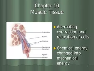
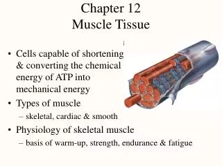
Introduction Composition: muscle cells, a little connective tissue. The cells have the property of contractility and are the functional units of muscle tissue. Myofiber: muscle fiber, because its elongated and thread-like shape, is termed. Sarcolemma: cell membrane is referred to as; Sarcoplasm: cytoplasm is termed; Sarcoplasmic reticulum: the smooth endoplasmic reticulum is called; Classification: skeletal (striated voluntary), cardiac (striated involuntary), and smooth muscles (unstriated involuntary). Voluntary, controled by somatic nerves; involuntary, by autonomic nerves
Skeletal muscle Cardiac muscle Smooth muscle
Skeletal Muscle Cell: long 4-40mm, 10-100μm in diameter, cylindrical; multinucleated- from the fusion of embryonic mononucleated myoblasts, nuclei are found at periphery, under sarcolemma; alternating dark and light bands (cross striations)
Myofibrils • Long, cylindrical in parallel • A band, I band, Z line, H band, M line • Sarcomere: • 1/2 I + A + 1/2 I ,shortest contractile unit of skeletal muscle.
2. 骨骼肌 Longitudinal Myofibrils Myofibril I band H Z A band Z M Sarcomere
A satellite cell (blue nucleus) adherent to a muscle fibre surface
Sarcomere M • Thick filaments • 1.6μm long and 15nm wide • Occupying A band • Made up of myosin molecules: like soybean sprouts, rods overlap; globular heads (halves of the beans) direct toward either of the ends forming cross bridgesandhave ATPase activity. • M line holds the thick filaments in place and bisects the H band. • Thick filaments overlap the thin filaments’ free ends.
Sarcomere M • Thin filaments • 1μm long and 8 nm wide; • One end is inserted into the Z line, the other is free and extends into the A band; • Composed of actin, tropomyosin and troponin(TnI, TnT, TnC).
Actin, monomers- 2 strands of globular (G-actin) monomers—twisted around each other in a double helical formation—F-actin polymers—filaments: αactinin anchors one end perpendicularly on the Z line. Each G-actin monomer contains a binding site for myosin. Tropomyosin, long (40 nm), thin molecules—2 polypeptide chains, bound head to tail—filaments: run over the actin subunits (7 subunits) alongside the outer edges of the groove between the two twisted actin strands.
Troponin: 3 subunits: TnT, tropomyosin binding site; TnC, calcium binding site; TnI, inhibits interaction between thin and thick filaments.
Arrangement • I band -- only thin filaments • A band --both thick and thin filaments • H band --only thick filaments • M line – fixation of thick filaments • Z line -- anchor for thin filaments
Transverse (T) tubules • Formed by sarcolemma invagination at A-I junctions, forming an anastomosingtubules surrounding every myofibril; • Responsible for rapid conduction of impulses.
Sarcoplasmic reticulum (L tubule) • Network of smooth endoplasmic reticulum • Encircling each myofibril between 2 adjacent T tubules. • Ends dilated and fused to form terminal cisternae
T tubule + 2 terminal cisternae = a triad. • Storing Ca2+, regulating concentration of Ca2+ within sarcoplasm.
Muscles: made up of skeletal muscle fibers surrounded by CT. F 1. Epimysium -- dense CT, surrounds entire muscle. 2. Perimysium -- fibrous sheath, surrounds muscle bundles. 3. Endomysium -- delicate loose CT, surrounds each muscle fibers. Ed Pe
Mechanism of Contractionsliding-filament hypothesis At the resting sarcomere, the binding site for myosin on the surface of actin is covered by tropomyosin. Ca2+ is released from the sarcoplasmic reticulum. Conformational change in the troponin complexes occurs, leading to movement of TnI away from the myosin head-binding sites on the actin filament. Binding of the myosin heads to the actin filaments activates ATPase in the heads. ATP is hydrolyzed into ADP and produce movement energy, resulting in cross-bridges twist and pulling the thin filament toward the center of the A band.
The neurotransmitter acetylcholine (ACh) is released from the synaptic vesicles of the axon terminal (1). ACh diffuses across the synaptic cleft and initiates the action potential on the sarcolemma (2). Depolarization of the sarcolemma and T tubules induces calcium release from the sarcoplasmic reticulum (3). Ca2+ binding to troponin leads to the tropomyosin shift (4) allowing for stronger myosin-actin binding and muscle contraction5-7. Muscle relaxation occurs when Ca2+is resequestered in the sarcoplasmic reticulum8.9.
Cardiac Muscle Pu General features Occurs exclusively in heart; More connective tissue and capillaries; Some specialized as Purkinje fibers.
Distinguishing features: Short cylindrical cells and branch at the ends. One or two nuclei per cell; located centrally, pale-staining perinuclear region. Actin and myosin filaments arranged in sarcomeres = striations. Myofibrils appear more branched than in skeletal muscle.
sarcolemma Z line Terminal cisterna Diad T tubule Sarcoplasmic reticulum A band I band H band Intercalated disc EM structure Larger T tubules at Z line level; Simple sarcoplasmic reticulum, small terminal cisternae; Diads are common consisting of T tubule and terminal cisternae on one side;
more sarcoplasm contains numerous mitochondria and glycogen particles;
Intercalated discs: Specialized cell junctions at Z lines Longitudinal portion has gap junction (G) providing synchronous contraction; Transverse portion has desmosomes(D)and fascia adherens(F, similar to zonula adherens)to enhance intercellular junction.
Smooth Muscle In the wall of blood vessels and hollow viscera, arranged in layers; Spindle in shape, with an oval, centrally located nucleus
B: Dense body P: Dense patch m: Myofilament Without striations,but contains thin and thick filaments;Adjacent cells are linked by gap junctions.
Myofilament Dense body Dense patch
Thin filaments are anchored by a-actinin to dense bodies and dense patches. Thick filaments have heads along most of their length and bare areas at the ends of the filaments, but they run in the opposite direction with the adjacent ones. The ratio of thin to thick filaments is 12:1. Bundles of myofilaments crisscross obliquely through the cell, forming a latticelike network. These bundles consist of thin filaments containing actin and tropomysin and thick filaments containing of myosin.
Thick filament Thin filament Thin filament Smooth muscle actin and myosin contract by a sliding filament mechanism similar to that which occurs in striated muscles. Smooth muscle fibers contain bundles of intermediate filaments that extend from one dense body to another, as well as to the dense patches, constituting a cytoskeleton.
The myosin filaments appear and the actin filaments are pulled toward and between them. Continued contraction involves the formation of more myosin filaments and further sliding of the actin filaments. The sliding actin filaments pull the attached dense bodies and dense patches closer together, shortening the cell. During relaxation, the myosin filaments disintegrate.