Download
1 / 16
170 likes | 390 Views
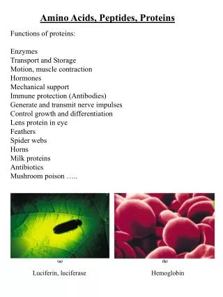
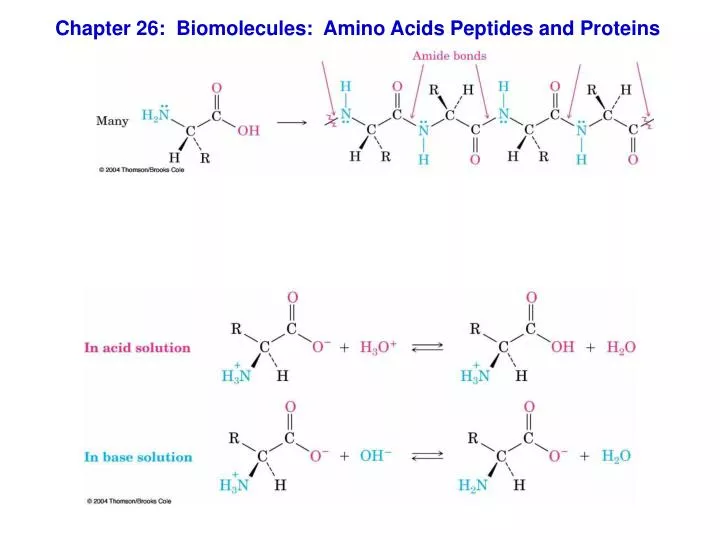
Chapter 26: Biomolecules: Amino Acids Peptides and Proteins. L-glyceraldehyde. D-glyceraldehyde. Chirality of Amino Acids. Glycine, 2-amino-acetic acid, is achiral In all the others, the carbons of the amino acids are centers of chirality

E N D
L-glyceraldehyde D-glyceraldehyde Chirality of Amino Acids • Glycine, 2-amino-acetic acid, is achiral • In all the others, the carbons of the amino acids are centers of chirality • The stereochemical reference for amino acids is the Fischer projection of L-serine • Proteins are derived exclusively from L-amino acids
Draw Fisher diagrams of L-Alanine (R = CH3) and L-cysteine (R = CH2-SH) and assign stereochemistry as R or S
Amino Acids: pKa and Isoelectric points
26.2 Isoelectric Points • In acidic solution, the carboxylate and amine are in their conjugate acid forms, an overall cation • In basic solution, the groups are in their base forms, an overall anion • In neutral solution cation and anion forms are present • This pH where the overall charge is 0 is the isoelectric point, pI
Titration Curves of Amino Acids • pKa’s determined from titration curve • If pKa values for an amino acid are known the fractions of each protonation state can be calculated (Henderson-Hasselbach Equation) • pH = pKa – log [A-]/[HA]
pI of any amino acid is the average of the two pKa’s that involve the neutral zwitterion
pI of any amino acid is the average of the two pKa’s that involve the neutral zwitterion • Contains an imidazole ring that is partially protonated in neutral solution • Only the pyridine-like, doubly bonded nitrogen in histidine is basic. • The pyrrole-like singly bonded nitrogen is nonbasic because its lone pair of electrons is part of the 6 electron aromatic imidazole ring (see Section 24.4). pI of Histidine ? (pKa’s = 1.82, 9.17, 6.00) Charge of Histidine at pH = 7.0?
Peptides (listed from N to C terminus) Draw structure of Ala-Ser-Asp. What is the net charge of the peptide at pH 7.5? Ala, R = CH3; Ser, R = CH2OH; Asp (R = CH2CO2H
26.10 Peptide Synthesis • Peptide synthesis requires that different amide bonds must be formed in a desired sequence • The growing chain is protected at the carboxyl terminal and added amino acids are N-protected • After peptide bond formation, N-protection is removed
Carboxyl Protecting Groups • Usually converted into methyl or benzyl esters • Removed by mild hydrolysis with aqueous NaOH • Benzyl esters are cleaved by catalytic hydrogenolysis of the weak benzylic C–O bond
Amino Group Protection • An amide that is less stable than the protein amide is formed and then cleaved after amide bond formed • The tert-butoxycarbonyl amide (BOC) protecting group is introduced with di-tert-butyl dicarbonate • Removed by brief treatment with trifluoroacetic acid
Peptide Coupling • Amides are formed by treating a mixture of an acid and amine with dicyclohexylcarbodiimide (DCC)
26.11 Automated Peptide Synthesis: The Merrifield Solid-Phase Technique
26.13 Protein Structure • The primary structure of a protein is simply the amino acid sequence. • The secondary structure of a protein describes how segments of the peptide backbone orient into a regular pattern. • The tertiary structure describes how the entire protein molecule coils into an overall three-dimensional shape. • The quaternary structure describes how different protein molecules come together to yield large aggregate structures