Download
1 / 54
590 likes | 1k Views
Cardiovascular System – Part I The Heart. Chapter 11 Kelly Trainor BIO 160. Objectives. Describe the location of the heart and identify its major anatomical areas Trace the pathway of blood through the heart Compare pulmonary and systemic circuits Explain the operation of heart valves

E N D
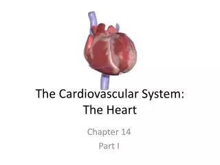
Cardiovascular System – Part IThe Heart Chapter 11 Kelly Trainor BIO 160
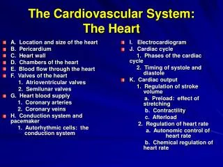
Objectives Describe the location of the heart and identify its major anatomical areas Trace the pathway of blood through the heart Compare pulmonary and systemic circuits Explain the operation of heart valves Name the elements of the intrinsic conduction system of the heart and describe the pathway of impulses through this system
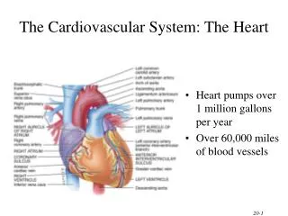
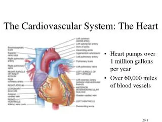
The Cardiovascular System • A closed system of the heart and blood vessels • The heart pumps blood • Blood vessels allow blood to circulate to all parts of the body • The function of the cardiovascular system is to deliver oxygen and nutrients and to remove carbon dioxide and other waste products
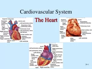
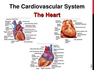
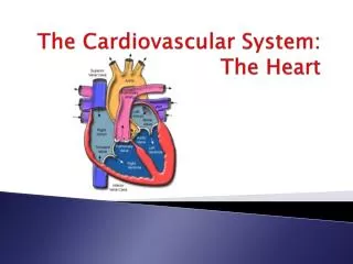
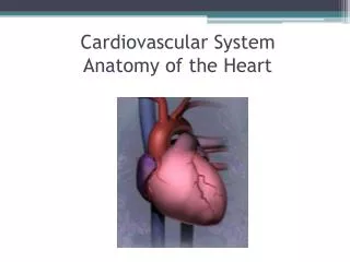
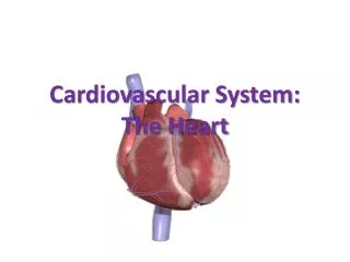
The Heart • Location • Thorax between the lungs in the inferior mediastinum • Orientation • Pointed apex directed toward left hip • Base points toward right shoulder • About the size of your fist
The Heart: Heart Wall • Three layers • Epicardium • Outside layer • This layer is the visceral pericardium • Connective tissue layer • Myocardium • Middle layer • Mostly cardiac muscle • Endocardium • Inner layer • Endothelium
The Heart: Chambers • Right and left side act as separate pumps • Four chambers • Atria • Receiving chambers • Right atrium • Left atrium • Ventricles • Discharging chambers • Right ventricle • Left ventricle
The Heart: Septa • Interventricular septum • Separates the two ventricles • Interatrial septum • Separates the two atria
The Heart: Chambers Figure 11.2c
The Heart: Valves • Allow blood to flow in only one direction to prevent backflow • Four valves • Atrioventricular (AV) valves—between atria and ventricles • Bicuspid (mitral) valve (left side of heart) • Tricuspid valve (right side of heart) • Semilunar valves—between ventricle and artery • Pulmonary semilunar valve • Aortic semilunar valve
The Heart: Valves Figure 11.2c
The Heart: Valves • AV valves • Anchored in place by chordae tendineae (“heart strings”) • Open during heart relaxation and closed during ventricular contraction • Semilunar valves • Closed during heart relaxation but open during ventricular contraction • Notice these valves operate opposite of one another to force a one-way path of blood through the heart
The Heart: Associated Great Vessels Figure 11.2c
Systemic and Pulmonary Circulations • Systemic circulation • Blood flows from the left side of the heart through the body tissues and back to the right side of the heart • Pulmonary circulation • Blood flows from the right side of the heart to the lungs and back to the left side of the heart
The Heart: Associated Great Vessels • Arteries • Aorta • Leaves left ventricle • Pulmonary arteries • Leave right ventricle • Veins • Superior and inferior venaecavae • Enter right atrium • Pulmonary veins (four) • Enter left atrium
The Heart: Associated Great Vessels Figure 11.2c
Blood Flow Through the Heart • Superior and inferior venaecavae dump blood into the right atrium • From right atrium, through the tricuspid valve, blood travels to the right ventricle • From the right ventricle, blood leaves the heart as it passes through the pulmonary semilunar valve into the pulmonary trunk • Pulmonary trunk splits into right and left pulmonary arteries that carry blood to the lungs
Blood Flow Through the Heart • Oxygen is picked up and carbon dioxide is dropped off by blood in the lungs • Oxygen-rich blood returns to the heart through the four pulmonary veins • Blood enters the left atrium and travels through the bicuspid valve into the left ventricle • From the left ventricle, blood leaves the heart via the aortic semilunar valve and aorta
The Heart: Conduction System • Intrinsic conduction system (nodal system) • Heart muscle cells contract, without nerve impulses, in a regular, continuous way • Special tissue sets the pace • Sinoatrial node = SA node (“pacemaker”), is in the right atrium • Atrioventricular node = AV node, is at the junction of the atria and ventricles • Atrioventricular bundle = AV bundle (bundle of His), is in the interventricular septum • Bundle branches are in the interventricular septum • Purkinje fibers spread within the ventricle wall muscles
Heart Contractions • Contraction is initiated by the sinoatrial node (SA node) • Sequential stimulation occurs at other autorhythmic cells • Force cardiac muscle depolarization in one direction—from atria to ventricles
Heart Contractions • Once SA node starts the heartbeat • Impulse spreads to the AV node • Then the atria contract • At the AV node, the impulse passes through the AV bundle, bundle branches, and Purkinje fibers • Blood is ejected from the ventricles to the aorta and pulmonary trunk as the ventricles contract
Heart Contractions • Tachycardia—rapid heart rate over 100 beats per minute • Bradycardia—slow heart rate less than 60 beats per minutes
Cardiovascular System – Part IIThe Blood vessels Chapter 11 Kelly Trainor BIO 160
Objectives Compare and contrast the structure and function of arteries, veins and capillaries Identify the body’s major arteries and veins and name the body region supplied by each Discuss the unique features of arterial circulation of the brain, and hepatic portal circulation
Blood Vessels: The Vascular System • Transport blood to the tissues and back • Carry blood away from the heart • Arteries • Arterioles • Exchanges between tissues and blood • Capillary beds • Return blood toward the heart • Venules • Veins
Blood Vessels: The Vascular System Figure 11.9b
Differences Between Blood Vessels • Walls of arteries are the thickest • Lumens of veins are larger • Larger veins have valves to prevent backflow • Skeletal muscle “milks” blood in veins toward the heart • Walls of capillaries are only one cell layer thick to allow for exchanges between blood and tissue Movement of Blood Through Vessels • Most arterial blood is pumped by the heart • Veins use the milking action of muscles to help move blood
Capillary Beds • Capillary beds consist of two types of vessels • Vascular shunt—vessel directly connecting an arteriole to a venule • True capillaries—exchange vessels • Oxygen and nutrients cross to cells • Carbon dioxide and metabolic waste products cross into blood
Major Arteries of System Circulation • Aorta • Largest artery in the body • Leaves from the left ventricle of the heart • Regions • Ascending aorta—leaves the left ventricle • Aortic arch—arches to the left • Thoracic aorta—travels downward through the thorax • Abdominal aorta—passes through the diaphragm into the abdominopelviccavity • Arterial branches of the ascending aorta • Right and left coronary arteries serve the heart
Major Veins of Systemic Circulation • Superior and inferior vena cava enter the right atrium of the heart • Superior vena cava drains the head and arms • Inferior vena cava drains the lower body
Arterial Supply of the Brain Figure 11.14
Hepatic Portal Circulation • Veins of hepatic portal circulation drain • Digestive organs • Spleen • Pancreas • Hepatic portal vein carries this blood to the liver • Liver helps maintain proper glucose, fat, and protein concentrations in blood • Major vessels of hepatic portal circulation • Inferior and superior mesenteric veins • Splenic vein • Left gastric vein
Hepatic Portal Circulation Figure 11.16
Cardiovascular System – Part IIIPhysiology of Circulation Chapter 11 Kelly Trainor BIO 160
Objectives Define blood pressure and pulse and name several pulse points List factors affecting and/or determining blood pressure Define hypertension and atherosclerosis, and describe possible health consequences of these conditions Describe the exchanges that occur across capillary walls
Pulse • Pulse • Pressure wave of blood • Monitored at “pressure points” in arteries where pulse is easily palpated • Pulse averages 70–76 beats per minute at rest
Blood Pressure • Measurements by health professionals are made on the pressure in large arteries • Systolic—pressure at the peak of ventricular contraction • Diastolic—pressure when ventricles relax • Write systolic pressure first and diastolic last (120/80 mm Hg) • Pressure in blood vessels decreases as distance from the heart increases