Download
1 / 39
390 likes | 481 Views
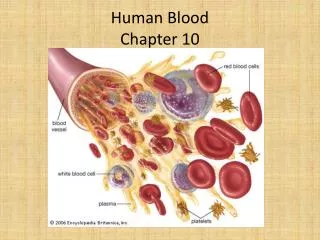
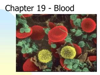
Blood Chapter 10. Blood. Connective tissue Consists of Formed elements and Plasma 45% - Formed elements 55% - Plasma Average total blood volume in adult male – 5-6 L Average total blood volume in adult female – 4-5 L Makes up about 8% of total body wt.

E N D
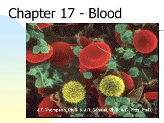
Blood Chapter 10
Blood • Connective tissue • Consists of Formed elements and Plasma • 45% - Formed elements • 55% - Plasma • Average total blood volume in adult male – 5-6 L • Average total blood volume in adult female – 4-5 L • Makes up about 8% of total body wt. • Oxygen-rich blood is scarlet red • Oxygen-poor blood is dull red • Heart pumps blood through blood vessels, which extend throughout the body
Blood • If blood is centrifuged • Erythrocytes sink to the bottom (45% of blood, a percentage known as the hematocrit) • Buffy coat contains leukocytes and platelets (less than 1% of blood) • Buffy coat is a thin, whitish layer between the erythrocytes and plasma • Plasma rises to the top (55% of blood)
Functions of Blood • Transport of gases, nutrients and waste products: eg. O2 • Transport of regulatory molecules: Transport hormones from endocrine organs to target organs • Regulation of pH : Buffers maintain the normal pH of most body tissues between 7.35 and 7.45 • Maintenance of body temperature: By absorbing and distributing heat throughout the body • Protection against foreign substances: e.g., antibodies, white blood cells • Clot formation: Protects excessive blood loss
Plasma • Liquid part of blood • Colloid – liquid suspension • Pale yellow fluid • Consists of 91% water, 7% protein, 2% other solutes, such as ions, gases, nutrients, waste products • Proteins • Albumins:58%, viscous • Maintain osmotic pressure • Globulins: 38%, transports lipids, carbohydrates, hormones, ions, involve in immunity (protection against microorganisms) • Fibrinogen: function in blood clotting • Serum: plasma without clotting factors
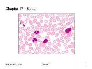
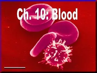
Formed Elements • Consists of red blood cell (95%), white blood cells and platelets (5%) • Red blood cells (erythrocytes): Contains hemoglobin, which colors the cell red, no nuclei • White blood cells (leukocytes): • Granulocytes:cytoplasm contains large granules; have multi-lobed nuclei. Three types: neutrophils, eosinophils, basophils • Agranulocytes:cytoplasm lack granules and nuclei that are not lobed. • Two types : lymphocytes and monocytes • Platelets (thrombocytes): Function in blood clotting
Red Blood Cells (Erythrocytes) • Biconcave disk • Increase surface area • Movement of gases faster • No nucleus • 5 million RBCs per cubic millimeter of blood • Components • 1/3 Hemoglobin – accounts • for red color • 2/3 Lipids, ATP, enzyme carbonic anhydrase
Functions of RBC • Transport oxygen from lungs to tissues • 98.5% of O2 attached to hemoglobin • 1.5% dissolved in plasma • Transport CO2 from tissues to lungs • 7% dissolved in plasma • 23% in combination with hemoglobin • 70% transported as bicarbonate ions • CO2 + H2O carbonic anhydrase H2CO3 H+ + HCO3-
Hemoglobin • Iron-containing protein • Binds strongly to oxygen • Each hemoglobin molecule has four oxygen binding sites • Each erythrocyte has 250 million hemoglobin molecules • Hemoglobin in one red blood cell transports 1 billion O2 molecules • Normal blood contains 12–18 g of hemoglobin per 100 mL blood
Hemocytoblaststem cells Lymphoidstem cells Myeloidstem cells Secondary stem cells Basophils Erythrocytes Platelets Eosinophils Lymphocytes Monocytes Neutrophils Production of Formed Elements • Process of blood cell production – Hematopoiesis • Occurs in red bone marrow of: • Ribs, sternum, vertebrae, pelvis, proximal femur and humerus • All formed elements derived from single population of stem cells – Hemocytoblasts • Hemocytoblasts divide into: • Lymphoid stem cell: Produces lymphocytes • Myeloid stem cell: Produce other classes of formed elements
Formation of Erythrocytes • Erythrocyte does not contain nuclei, cannot divide, grow, or synthesize proteins • Wear out in 100 to 120 days • When worn out, RBCs are eliminated by phagocytes in the spleen or liver • Lost cells are replaced by division of hemocytoblasts to new RBCs in the red bone marrow
Control of Erythrocyte Production • Rate of erythrocyte productions is controlled by a hormone (erythropoietin) • Kidneys produce most erythropoietin as a response to reduced oxygen levels in the blood • Homeostasis is maintained by negative feedback from blood oxygen levels
Erythrocyte Disorders • Anemia – Deficiency of hemoglobin in blood • Reduces blood`s ability to transport oxygen • Unable to support normal metabolism • Symptoms: fatigue, paleness, shortness of breath, and chills • Polycythemia - • Is an excessive or abnormal increase in the number of erythrocytes
Erythrocyte Disorders • Sickle-cell anemia – RBCs become sickle shape • single mutation changes amino acid in globin of Hb • causes RBCs to become sickle-shaped
White Blood Cells (Leukocytes) • Make up 1% of the total blood volume • 4,000 to 11,000 WBC per cubic millimeter of blood • No hemoglobin • Contains nuclei • Protect body against microorganisms • Remove dead cells and debris • Motile • Movements: • Ameboid • Diapedesis: Enter tissue by diapedesis, cells become thin, elongate and move between blood capillaries • Chemotaxis: move toward foreign materials or damaged cells • Phagocytize bacteria and dead cells
White Blood Cells (Leukocytes) • Two major categories of Leukocytes: • Granulocytes: • Contains cytoplasmic granules • Lobed nuclei • Neutrophils • Eosinophils • Basophils • Agranulocytes: • Lack granules • Spherical or kidney shaped nuclei • Lymphocytes • Monocytes
White Blood Cells (Leukocytes) • Neutrophils: • Comprise 60-70% of white blood cells • Contains small cytoplasmic granules • multilobed nucleus • Phagocytize bacteria, foreign matter • Eosinophils: • Comprise 2-4% of white blood cells • Their no. increases during allergies and infections by parasitic worms • Destroy parasitic worms: tapeworms, flukes, pinworms, hookworms
White Blood Cells (Leukocytes) • Basophils: • Comprise 0.5-1% of white blood cells • Increases no. in both inflammation & allergies • Release heparin, which inhibits blood clotting • Lymphocytes: • Comprise 20-25% of white blood cells, smallest WBCs • Nucleus fills most of the cell • Produced in red bone marrow, migrate to lymphatic tissues • Play role in immunity, produce antibodies • destroy tumor cells
White Blood Cells (Leukocytes) • Monocytes: • Comprise 3-8% of white blood cells, largest WBCs • leave circulation and become macrophages • Phagocytize bacteria, dead cells, debris
Formation of White Blood Cells and Platelets • Controlled by hormones • Colony stimulating factors (CSFs) and interleukins prompt bone marrow to generate leukocytes • Thrombopoietin stimulates production of platelets
Leukocyte Disorders • Abnormal numbers of leukocytes • Leukocytosis • WBC count above 11,000 leukocytes/mm3 • Generally indicates an infection • Including viral, bacterial, fungal, or parasitic infection, cancer, hemorrhage • Leukopenia • Abnormally low leukocyte level • Commonly caused by certain drugs such as corticosteroids and anticancer agents • Other causes are: malaria, HIV, tuberculosis
Leukocytes disorders: Leukemia • Cancer of red bone marrow • Abnormal production of WBCs • WBCs are immature • Lack normal immunologic function • Interferes with RBCs and platelet production • Lead to anemia, bleeding • Death is caused by internal hemorrhage and infections • Treatments include radiation, antileukemic drugs, and • bone marrow transplants
Platelets (Thrombocytes) • Small fragments of cells • Produce within red marrow • Derive from megakaryocytes • Cell fragments pinched off from megakaryocytes in red bone marrow • Essential for blood clotting • Prevent blood loss • Normal platelet count = 300,000/mm3
Hemostasis • Series of reactions for stoppage of bleeding • Three phases occur in rapid sequence • Vascular spasms • Platelet plug formation • Coagulation (blood clotting) • Vascular spasms: • Immediate constriction of blood vessel - injury • Close small vessels • Stop blood flow
Hemostasis • Platelet plug formation • Collagen fibers are exposed by a break in a blood vessel • Platelets become “sticky” and cling to fibers • Anchored platelets release chemicals to attract more platelets • Platelets pile up to form a platelet plug
Hemostasis • Coagulation • Injured tissues release tissue factor (TF) • PF3 (phospholipid that coats the surface of platelets) interacts with TF, blood protein clotting factors, and calcium ions to trigger a clotting cascade • Prothrombin activator converts prothrombin to thrombin (an enzyme) • Thrombin joins fibrinogen proteins into hair-like molecules of insoluble fibrin • Fibrin forms a meshwork (the basis for a clot)
Hemostasis • Blood usually clots within 3 to 6 minutes • Endothelium regenerates • And the clot is broken down after tissue repair
Disorders of Hemostasis • Thrombus • A clot in an unbroken blood vessel • Can be deadly in areas like the heart • Embolus • A thrombus that breaks away and floats freely in the bloodstream • Can later clog vessels in critical areas such as the brain
Disorders of Hemostasis • Thrombocytopenia • Platelet deficiency • Even normal movements can cause bleeding from small blood vessels that require platelets for clotting • Hemophilia • Hereditary bleeding disorder • Normal clotting factors are missing
Blood Groups and Transfusions • Large losses of blood have serious consequences • Loss of 15–30% causes weakness • Loss of over 30% causes shock, which can be fatal • Transfusions are the only way to replace blood quickly • Transfused blood must be of the same blood group
Blood Grouping • Surface of RBC have specific glycoprotein - antigens • These antigens are: • Unique to the individual • Antibodies(present in plasma) can bind to specific RBC antigens • If different blood cell type is transfused, antigen combines with antibody – agglutination (clumping) takes place • Antigens – agglutinogens • Antibodies - agglutinins
ABO Blood Groups • Based on the presence or absence of two antigens • Type A • Type B • The lack of these antigens is called type O
ABO Blood Groups • The presence of both antigens A and B is called type AB • The presence of antigen A is called type A • The presence of antigen B is called type B • The lack of both antigens A and B is called type O
ABO Blood Groups • Blood type AB can receive A, B, AB, and O blood • Universal recipient • Blood type B can receive B and O blood • Blood type A can receive A and O blood • Blood type O can receive O blood • Universal donor
Rh Blood Groups • First studied in rhesus monkeys • Types • Rh positive: Have D antigen present on surface of RBCs • Rh negative: Do not have D antigen • Most Americans are Rh+ (Rh positive) • Problems can occur in mixing Rh+ blood into a body with Rh– (Rh negative) blood
Rh Dangers During Pregnancy • Danger occurs only when the mother is Rh– and the father is Rh+, and the child inherits the Rh+ factor • RhoGAM shot can prevent buildup of anti-Rh+ antibodies in mother’s blood
Rh Dangers During Pregnancy • The mismatch of an Rh– mother carrying an Rh+ baby can cause problems for the unborn child • The first pregnancy usually proceeds without problems • The immune system is sensitized after the first pregnancy • In a second pregnancy, the mother’s immune system produces antibodies to attack the Rh+ blood (hemolytic disease of the newborn)
ERYTHROBLASTOSIS FETALIS • Rh+ fetus / Rh- mother • Fetal organ damage FIRST PREGNANCY SECOND PREGNANCY maternal circulation (Rh-) (RH-) antibodies develop in the mother after delivery fetal circulation (Rh+) Agglutination of fetal (RH+) fetus