Download

1 / 21
240 likes | 528 Views
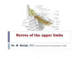
David Fu. The Bones of Upper Limbs Medical College of Jiujiang University. The Bones of Limbs.

E N D
David Fu The Bones of Upper LimbsMedical CollegeofJiujiang University
The Bones of Limbs The limbs articulate with the trunk by the should girdle and pelvic girdle respectively. The upper limb is characterized by considerable mobility and is adapted for grasping and manipulating. The lower limb is specialized for locomotion, bearing weight and maintaining equilibrium, so for same reason, the bones of lower limb are more massive than those of the upper limb.
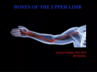
Bones of upper limbs Composition: 32×2=64 • Should girdle: clavicle,scapula • Bones of free upper limb: • Humerus in arm • Radius and ulna in forearm • Carpal, metacarpalsand phalanges in hand
Shoulder (Pectoral) Girdle • Consists of scapula and clavicle • Clavicle articulates with sternum (sternoclavicular joint) • Clavicle articulates with scapula (acromioclavicular joint) • Scapula held in place by muscle only • Upper limb attached to shoulder girdle at shoulder (glenohumeral joint)
Clavicle (collarbone) • The clavicles lies horizontally in the anterolateral to the superior aperture of thorax. • It is a long bone with one shaft and two ends. • S-shaped bone with two curves • medial 2/3 curve convex anteriorly/lateral 1/3 concave anteriorly • Sternal end medially and acromial end laterally • Extends from sternum to scapula above 1st rib
Clavicle • Fracture site is junction of curves • Ligaments attached to clavicle stabilize its position.
Scapula • The scapula lies on the posterolateral aspect of the thorax, overlying the 2nd to 7th ribs. • It is a flat, triangular bone, with two surfaces, three borders and three angles. • Two surfaces • Anterior surface concave: subscapular fossa • Posterior surface: supraspinousfossae and infraspinous fossae,spine of scapula, acromion • Three borders • Superior: coracoid process scapular notch • Lateral (axillary) border • Medial (vertebral) border • Three angles • Superior: opposite to the 2nd rib • Inferior: opposite to the 7th rib or 7th intercostals space • Lateral: glenoid cavity supraglenoid tubercles and infraglenoid tubercles
Anterior Surface of Scapula • Subscapular fossa filled with muscle • Coracoid process for muscle attachment
Posterior Surface of Scapula • Scapular spine ends as acromion process • a sharp ridge widening to a flat process • Glenoid cavity forms shoulder joint with head of humerus • Supraspinous & infraspinous fossa for muscular attachments
Bones of Free Upper Limb • Each free upper limb = 30 bones • humerus within the arm • ulna & radius within the forearm • carpal bones within the wrist • metacarpal bones within the palm • phalanges in the fingers • Joints • shoulder (glenohumeral), elbow, wrist, metacarpophalangeal, interphalangeal
Humerus --- Proximal End • It is the longest and largest bone of upper limb. It is divided into a shaft and two ends. • Part of shoulder joint • Head & anatomical neck • Greater & lesser tubercles for muscle attachments • Intertubercular sulcus or bicipital groove • Surgical neck is fracture site • Shaft is almost cylindrical in the upper half, prismatic and flattened below. About the middle of body, the lateral border forms a rough, triangular elevation, the deltoid tuberosity for insertion of the deltoid.
On the posterior surface of the shaft there is an oblique groove, the sulcus for radial nerve which directed obliquely from superior-medial to inferior-lateral and transmitted the radial nerve and deep brachial artery. The fracture of middle part of shaft can damage the radial nerve.
Humerus --- Distal End • Forms elbow joint with ulna and radius • Capitulum articulates with head of radius • Trochlea • articulation with trochlear notch of ulna • Olecranon fossa • posterior depression for olecranon of ulna • Medial & lateral epicondyles • attachment of forearm muscles • The ulnar nerve runs downwards in the sulcus for ulnar nerve on the back of medial epicondyle.
Humerus • Upper end: head of humerus,anatomical neck, greater and lesser tubercles, crests of greaterand lesser tubercle, intertubercular groove, surgical neck • Shaft: deltoid tuberosityon lateral surface, and a groove for radial nerve on posterior surface • Lower end: lateral and medial epicondyles, capitulum, trochlea, coronoid fossa and radial fossa (anteriorly) and olecranon fossa (posteriorly), and sulcus for ulnar nerve
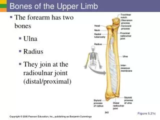
Ulna & Radius --- Proximal End • Ulna (on little finger side) • trochlear notch articulates withhumerus & radial notch with radius • olecranon process forms joint of elbow • Radius (on thumb side) • head articulates with capitulum of humerus & radial notch of ulna • tuberosity for muscle attachment
Elbow Joint • Articulation of humerus with ulna and radius • Ulna articulates with trochlea of humerus • Radius articulates with capitulum of humerus • Interosseous membrane between ulna & radius provides site for muscle attachment
Ulna and Radius - Distal End • Ulna --styloid process • head separated from wrist joint by fibrocartilage disc • Radius • forms wrist joint with scaphoid, lunate & triquetrum • forms distal radioulnar joint with head of ulna
Radius • Upper end: head of radius, neck of radius, radial tuberosity, and articular circumference • Shaft:interosseous border • Lower end: styloid process laterally, ulnar notch medially, and carpal articular surface inferiorly
Ulnar • Upper end: olecranon coronoid process trochlear notch radial notch ulnar tubersity • Lower end styloid process head of ulna
8 Carpal Bones (wrist) • Proximal row - lateral to medial • scaphoid - boat shaped • lunate - moon shaped • triquetrum - 3 corners • pisiform - pea shaped • Distal row - lateral to medial • trapezium - four sided • trapezoid - four sided • capitate - large head • hamate - hooked process • Carpal tunnel--tunnel of bone & flexor retinaculum • She looks too pretty, try to catch her.
Metacarpals and Phalanges • Metacarpals • 5 total----#1 proximal to thumb • base, shaft, head • knuckles (metacarpophalangeal joints) • Phalanges • 14 total: each is called phalanx • proximal, middle, distal on each finger, except thumb • base, shaft, head