Download
1 / 25
260 likes | 680 Views
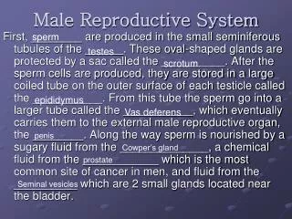
Male reproductive system. three accessory organs seminal vesicle prostate bulbourethral glands (Cowper glands) reproductive ducts vas deferens (ductus deferens) urethra. -vas deferens: conducting tube from testis to urethra

E N D
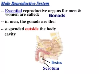
Male reproductive system • three accessory organs • seminal vesicle • prostate • bulbourethral glands (Cowper glands) • reproductive ducts • vas deferens (ductus deferens) • urethra
-vas deferens: conducting tube from testis to urethra -epididymus – connection between the testis and the vas deferens -stores immature sperm -vas deferens + blood vessels + nerves + lymphatics = spermatic cord (passes through inguinal canal) -urethra: 3 sections: A. prostatic - runs through prostate B. membranous - between prostate and penis C. spongy - through penis
Reproductive glands Bladder Rectum • seminal vesicles, prostate, bulbourethral glands • -produce fluid that combine with sperm to make semen • -semen: alkaline, activates sperm cells • 1. prostate: surrounds the urethra • -secretes a thin, milky fluid that enhances sperm motility and neutralizes vaginal fluid • 2. seminal vesicles: connect to urethra via the ejaculatory ducts • -secretes an alkaline fluid that contains sugars and prostaglandins (stimulates uterine contractions) • 3. bulbourethral glands: 2 glands behind the prostate • -secrete a fluid that lubricates the penis Prostate Corpus spongiosum Prostatic Urethra Membranous Urethra Corpus cavernosum Spongy/ Penile Urethra Glans Penis Testes External Urethral Orifice
Bladder Rectum Prostate Corpus spongiosum Prostatic Urethra Membranous Urethra Corpus cavernosum Spongy/ Penile Urethra Glans Penis Testes External Urethral Orifice
scrotum = supportive structure for the testes • hangs from the root of the penis • externally- single pouch separated at the midline by a raphe • internally – divided by a scrotal septum into two sacs each containing 1 testis • the scrotum contains two muscles • dartos muscle (smooth muscle) & the cremaster muscle – skeletal muscle that is a continuation of the internal oblique -testes within the sac is fed by branches off the testicular artery and drained by a series of veins called the pampiniform plexus
pampiniform plexus Vas deferens testicular artery testicular artery branches Epididymus Seminiferous Tubules Tunica Albuginea Tunica Vaginalis -testis: develop internally near the kidneys and descend during the latter half of the seventh month gestation -covered by two protective membranes: 1. tunica vaginalis –derived from the peritoneum 2. tunica albuginea – internal to the TV -extends inward to divide the testes into lobules (200-300) -each lobule contains 1 to 3 coiled seminiferous tubules for sperm production - lined with spermatogenic cells that produce sperm -contain sperm stem cells = spermatogonium
-testis: also contain two additional cell types: • -Sertoli cells = sustenacular cells • -found in the seminiferous tubule • -secretion of growth factors that mediate spermatogenesis • - consume the unneeded portions of the spermatazoa as they mature • - create a privileged immune environment – prevents autoimmune reactions • -interstitial cells – located between the seminiferous tubules • -for testosterone production
Spermatogenesis • sperm development – from sperm stem cells called spermatogonium • these spermatogonium develop in the embryonic testes • the spermatogonium remain dormant in the testes until puberty • mature sperm cells = spermatozoa • takes 60-75 days to complete • 1. differentiation of the spermatogonium into primary spermatocytes (2n) • 2. start of meiosis • 3. formation of secondary spermatocytes (n) – 23 chromosomes comprised of two chromatids • 4. completion of meiosis and formation of four spermatids (23 chromosomes each made up of one chromatid) • 5. spermiogenesis – development of spermatids into a sperm cell (head, acrosome and flagella development)
Sperm • 300 million made each day • 60 um long • major parts • 1. head: contains the nucleus with 23 highly condensed chromosomes (one chromatid) • 2. acrosome: covers the anterior 2/3 of the head • contains digestive enzymes to dissolve the protective barriers of the egg (hyaluronidase and proteases) • 3. tail or flagellum • neck - constricted region just behind the head • contains centrioles for the production of the microtubules for the tail • middle piece – contains mitochondria arranged in a spiral • principal piece – longest portion of the tail • end piece – terminal portion of the tail end piece principal piece
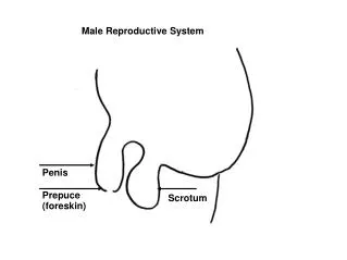
made up of a: • root • body (or shaft) • glans -conveys urine and semen -body is found externally, root is found internally (attached to the pubic ramus) -body is comprised of two tissue types: erectile tissue surrounded by connective tissue A. corpus cavernosum – larger spaces B. corpus spongiosum - smaller spaces & surrounds the urethra -corpus spongiosum enlargens at the tip - glans penis (sensory receptors) -glans penis covered with a loose fold of skin = prepuce
Corpus cavernosum Corpus spongiosum Spongy/ Penile Urethra Vas Deferens Epididymus Spermatic Cord Testes
-release of Gonadotropic releasing hormone (GnRH) from the hypothalamus • which stimulates the anterior pituitary gland • -anterior pituitary releases 2 gonadotropins (FSH and LH) • Follicle stimulating hormone - stimulates spermatogenesis • -also stimulates the production of the hormoneinhibin– inhibits GnRH • 2. Leutinizing hormone - stimulates male hormone production • -male hormones: androgens (e.g. testosterone) • -testosterone: secondary sex characteristics, reproductive organ development and • maintenance • -rise in testosterone causes a negative feedback system: hypothalamus is inhibited
Testosterone • two major forms: testosterone and dihydroxytestosterone (DHT) • testosterone and DHT both bind to same receptors • targets – bone, muscle (androgenic) • effects • 1. stimulates the male pattern of the reproductive system • testosterone stimulates development of the epididymus, vas deferens, ejaculatory duct and seminal vesicle • DHT derivative stimulates development of external genitalia • testosterone is converted in the brain to estrogens – development of certain brain regions in males • 2. development of male sexual characteristics • 3. development of sexual function • male sexual behavior • spermatogenesis • libido in both males and females • females – androgen release by the adrenal cortex • 4. stimulation of anabolism • stimulate protein synthesis
Development of male reproductive structures • Two embryonic ducts within the developing fetus • Mullerian ducts – female • Wolffian ducts - male • Role of the Sertoli cells • Presence of the SRY gene • Gene product induces differentiation of the Sertoli cells • Production of anti-Mullerian hormone • Role of the Leydig cells • Testosterone is critical for development of internal reproductive structures • Also required for development of external genitalia
-uterus: receives and nourishes the embryo -comprised of a body, a curved portion (fundus) and the cervix -uterine wall outer perimetrium, muscular myometrium and inner endometrium -endometrium: mucosal layer covered with epithelium -rich blood supply, sloughed off during menstruation -comprised of a deeper basal layer and an outer functional layer
-uterine tubes or oviducts (Fallopian tubes): conduction of egg from ovary to uterus -expands at end near the ovary = infundibulum with fimbrae (fingers) for the “catching” of the released egg -fertilization occurs in the uterine tubes -cervix: projects into the vaginal canal -vaginal canal broadens to surround the cervix – fornix -site for the insertion of the penis and deposition of sperm -opens at the exterior as the vulva (female external genitalia)
Round ligament Broad ligament Fundus Fundus Ovary Bladder Fornix Body Fimbrae of oviduct Fallopian Tube (oviduct) Cervix Bladder Rectum Vaginal canal Urethra External urethral orifice Labia minora Vaginal orifice Labia majora
-uterus is associated with a round ligament and a broad ligament -round ligament originates near the fallopian tubes – runs through the female inguinal canal to insert onto the labia majora/mons pubis -broad ligament – wide fold of peritoneum -connects the perimetrium of the uterus to the walls and floor of the pelvis -part of the broad ligament is the mesovarium – covers the ovary -uterus is fed by the uterine artery
-known as the vulva -mons pubis – mass of adipose tissue that lays on top of the pubic bone – protects the pubic bone during intercourse -mons pubis divides into the labia majora and minora -labia majora: folds of adipose tissue that enclose and protect the labia minora -labia minora: surrounds the external urethral orifice and the vaginal orifice -converge anteriorly to form a hood over the clitoris -clitoris: erectile tissue with a glans, body and a root
-ovary: production of oocytes -inner medulla and outer cortex -medulla - connective tissue with blood & lymphatic vessels and nerves -cortex - granular tissue due to the presence of tiny ovarian follicles containing egg stem cells (oogonium)
-ovarian follicles mature and contain the developing oocyte = oogenesis • growth of primary follicles into secondary follicles with the primary oocyte • -primary oocyte splits in the follicle unequally producing: • 1. a large secondary oocyte - released during ovulation 2. a smaller polar body • 2. secondary follicle matures -> vesicular /tertiary follicle with a large secondary oocyte (for ovulation) • 3. ruptured follicle degenerates into a corpus hemorrhagicumand then into a corpus luteum - hormone production
Oogenesis • begins before birth • early fetal development – primordial germ cells in the developing ovaries differentiate to form oogonia (egg stem cells) • at birth meiosis in the germ cells results in the development of primary follicles containing primary oocytes – stopped at prophase I of meiosis • at puberty - release of FSH and LH each month causes the development of a primary oocyte into a secondary oocyte followed by ovulation • coincides with the development of the primary follicle, (primary oocyte) into a secondary follicle (primary oocyte) and finally into a tertiary follicle (secondary oocyte) • tertiary follicle – ovulation of secondary oocyte • fertilization is required for the completion of meiosis II • therefore during oogenesis, ONE oocyte merges with the sperm and THREE polar bodies form
-GnRH causes release of FSH and LH from anterior pituitary -FSH causes maturation of follicles -LH results in development of corpus luteum -corpus luteum produces and releases estrogen and progesterone -estrogen and progesterone: regulate pregnancy, menstruation, secondary sex characteristics & development of sex organs at puberty
-divided into two cycles 1. uterine cycle 2. ovarian cycle -Ovarian cycle: -rise in FSH matures the follicle -follicle begins to produce follicular estrogen (estradiol) (Day 9) -gradual increase in LH and a decrease of FSH -sudden increase in FSH and LH plus follicular estrogen (Day 14) coincides with ovulation -the increase in follicular estrogen is what causes the spike in LH -ovulation = LH spike is the trigger (Day 14) -drop in LH, FSH and follicular estrogen after ovulation -but FSH will begin to climb at the end of the cycle
-Uterine cycle: – build up of the endometrium followed by menstruation -menstrual cycle initiated by FSH (Day 1) -rise in follicular estrogen starts thickening of uterine lining = known as the proliferative phase -increased secretion of luteal estrogens + synthesis of progesterone by the corpus luteum increases blood content of uterine lining to thicken it = known as the secretory phase -ovulation marks the change from proliferative to secretory phase -estrogen and progesterone begin to drop about day 25 to 28 - disintegration of uterine lining (stratum functionalis) -Menstruation (3 to 7 days)