Download
1 / 34
340 likes | 526 Views
Inelastic X-Ray Scattering Studies of Phonon Propagation and Damping in Biomolecular Assemblies. Sow-Hsin Chen (sowhsin@mit.edu). Department of Nuclear Science and Engineering, MIT, Cambridge, MA, USA. Collaborators: Y. Liu (MIT), D. Liu (MIT), D. Berti (U, Florence), E. Fratini (U, Florence),

E N D
Inelastic X-Ray Scattering Studies of Phonon Propagation and Damping in Biomolecular Assemblies Sow-Hsin Chen (sowhsin@mit.edu) Department of Nuclear Science and Engineering, MIT, Cambridge, MA, USA Collaborators: Y. Liu (MIT), D. Liu (MIT), D. Berti (U, Florence), E. Fratini (U, Florence), P. Baglioni (U, Florence), H. Sinn (APS), A. Alatas (APS), A. Said (APS), E. Alp (APS) Research supported by Material Sciences Program, BES, US DOE An invited lecture given at NSLS-ll Workshop on IXS, BNL, Feb. 7th-8th, 2008
1. Thomson scattering of x-rays from atomic electrons is a coherent scattering, irrespectively of atomic composition of a bio-molecule. There is no need to deuterate the bio-molecule as in a coherent inelastic neutron scattering experiment. While the number of x-ray photons or neutrons at the sample are comparable ( 3108 photons or neutrons per second), a synchrotron x-ray beam size is much smaller (200 m 100 m) than that in an INS spectrometer. In practice, for a 40 wt% calf-thymus Na-DNA sample in the hexagonal liquid crystalline phase, a film of dimension 0.2 mm (thick) 0.2 mm (wide) 10 mm (long) on a quartz plate is sufficient, which is of order of 0.4 mg of DNA. Ordered 40 wt% calf-thymus Na-DNA samples of thickness 0.2 mm have been used successfully by us in APS in the past years. Advantages of Inelastic X-Ray Scattering for Investigation of Collective Modes in Bio-Materials
3. Even if one deuterates a bio-molecule, the dynamic structure factor as measured in an INS experiment is a weighted sum of contributions from partial structure factors of deuterium, carbon, oxygen, nitrogen and phosphor, etc.. Fortunately, the dynamic structure factor measured in an IXS is practically dominated by the phosphate-phosphate PSF coming from the backbones of the DNA molecules. So the interpretation of the nature of the collective modes measured is simpler. 4. An IXS spectrum measured with an energy resolution of E = 0.1 meV can be calculated from the corresponding intermediate scattering function (ISF) computed with a time window of 15 ps. Modern MD simulations of bio-macromolecules are routinely pursued up to 100 ps. Thus measured IXS spectra of bio-macromolecules can be used to critically test the force field models of proteins, lipid bilayers and DNA’s. Advantages of Inelastic X-Ray Scattering for Investigation of Collective Dynamics in Bio-Materials
Three Effective Eigenmode (TEE) Model The dynamic structure factor is given as, with and zu(k),fuT(k),zT(k) all real numbers. Hydrodynamic limit (k0): The three eigenvalues, to the order of O(k2), are: where is the sound damping.
For finite k, zu(k),fuT(k),zT(k) become arbitary functions of k. However, in most cases, the eigenvalues of the matrix H consist of one real number zh and a couple of conjugate complex numbers s± is. One can therefore write the DSF in general in the hydrodynamic-like form: Damped Harmonic Oscillator limit (fuT(k)=0) The amplitude of the central peak of S(k,) is zero, the side peaks can be written as : The practical DHO model
Generalized Dynamic Structure Factor for Polyatomic Molecules The Dynamic Structure Factor can be written as a weighted sum of the partial dynamic structure factor : where is the atomic form factor of the -species of atoms . S(k,) is given by GTEE model. The structure factor measured by an X-rays diffraction experiment is thus where the partial structure factor is given by The second moment sum rule can be shown to be
SUMMARY – (1) • Geralized Three Effective Eigenmode Theory is an appropriate theory for analyses of IXS spectra from lipid bilayers. It allows one to extract: the dynamic structure factor, the phonon dispersion relation, the phonon damping, and thermal diffusion coefficients of the bilayers. • The phonon dispersion relation of a bilayer shows a deep minimum at the peak of the structure factor. This minimum frequency is lower in the LC phase than that in the gel phase. • Given a chain length, the high-frequency sound speed is higher in the gel phase than in the LC phase. For DMPC it is 2230 m/s vs 1936 m/s.
SUMMARY – (2) • The phonon frequency at the peak of the dispersion relation increases with the chain length while that at the valley decreases with the chain length. • Addition of cholesterol to DMPC, originally in the LC phase, has an effect of changing the dynamic structure factor and the phonon dispersion relation closer to that of the gel phase. This can be interpreted to mean that an addition of cholesterol to bilayer at LC phase temperature has a tendency to increase the order of lipid molecules in the bilayer and makes the bilayer more solid-like. • Addition of cholesterol to a bilayer hardly changes the thermal diffusion coefficient DT of the bilayer.
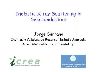
DNA: A molecular model: 1 hydrogen, 2 oxygen, 3 carbon in the helical phosphate ester chains, 4 carbon and nitrogen in the cross-linked purine and pyrimidine bases, 5 phosphorus; B double helix
F. Livolant, A. Leforestier, Progress in Polymer Science, 21, 1115, 1996 linear charge spacing between phosphate groups along DNA axis D = 1.7 Ǻ
Important Effects of Counter-Ion Atmosphere on Properties of DNA • Counter-Ion Condensation. • Counter-ions neutralize DNA molecules and cause DNA to aggregate. • Counter-ions can cause charge inversion of DNA molecules, which • introduces DNA aggregation even in dilute solutions. • 2.DNA Structural Stability. • Alkaline earth metal ions can stabilize B-DNA against thermal denaturation. • Transition metal ions preferentially bind to the nucleic bases, • disrupting hydrogen bonding between base pairs and destabilizing • the B form DNA. • 3.DNA Conformational Change. • Mg2+ can influence B-Z conformation transition; DNA A-B transition • is also dependent on counter-ions.
N = counterion valency, (the distance between elementary charges which the electrostatic interaction energy equals the thermal energy) Theory of Counterion Condensation (G. S. Manning, The molecular theory of polyelectrolyte solutions with applications to the electrostatic properties of polynucleotides, Quartely Reviews of Biophysics II, 2, 179, 1978 ) In a linear polyelectrolyte of sufficiently high charge density, a certain fraction of its charge is neutralized due to the territorial binding of counterions in the immediate environment. The condensed counterions are free to diffuse along the polymer axis, but inhibited from diffusing away radially. The fraction of groups neutralized by the counterion condensation, b = linear charge spacing between phosphate groups along DNA axis = 1.7 Ǻ
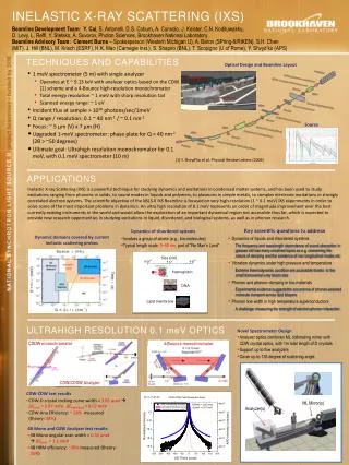
The Intermediate Scattering Function, F(Q,t) 40 wt% Na-DNA in H2O 40 wt% Na-DNA in 0.083 M CaCl2
Summary 1. Generalized Three Effective Eigenmode Theory is an appropriate theory for analyses of IXS spectra of DNA samples. It allows one to extract: the dynamic structure factor, the phonon dispersion relation, the phonon damping and the relaxation rate of the central peak. 2. The phonon dispersion relation of 40 wt% calf-thymus Na-DNA in water is constructed. At low Q, it is linearly dependent on Q with the sound speed similar to that of water (about 3100 m/s). It shows an oscillatory behavior in Q range of 2 nm-1 to 30 nm-1, similar to that obtained from lipid bilayers, and it extends to a considerably higher Q than that observed in molecular liquids and liquid metals. 3. In the interval of 10 nm-1 < Q < 22.5 nm-1, the phonon peaks in the 40 wt% calf-thymus Na-DNA in solutions with divalent and trivalent counter-ions are completely dampened out. We conjecture that this is due to an increased phonon damping caused by the increased multivalent counter-ion condensation, which change the water mobility around DNA. This leads to a concept of the “counter-ion friction”.
Calculated and Measured Average Structure Factors of Globular Proteins P. Etchegoin, Glassylike low-frequency dynamics of globular proteins,PRE, 58, 845, (1998) Intermediate range order of 2-6 Ǻ Structure factor of Lysozyme measured by x-ray scattering
Phonon Energies of Lysozyme and BSA Below and Above the Glass Transition Temperature
The Structural Effect on Phonon Dispersion in Lysozyme and BSA
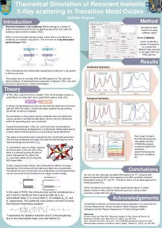
Phonon Dispersion and Brillouin-to-central-peak Intensity Ratio of Lysozyme and BSA D. Liu and S.-H. Chen, et al, to be published
Preliminary Observation of Phonons in Globular Proteins We analyzed the IXS spectra by the practical DHO model. The agreement with experimental data is satisfactory. All globular proteins have a universal average structure factor showing a peak at Q = 15 nm-1. Phonons can only be observed for Q values at and larger than this peak. The peak phonon energy changes significantly at the temperature of the so-called glass transition of protein at Tg = 225 K.
References: • C. Y. Liao, S. H. Chen, and F. Sette, “Analysis of inelastic X-ray scattering spectra of low temperature water,” Phys. Rev. E, 61:1518, 2000 • C. Y. Liao, and S. H. Chen, “Theory of the generalized dynamic structure factor of polyatomic molecular fluids measured by inelastic x-ray scattering,” Phys. Rev. E, 64:021205, 2001 • S. H. Chen, C. Y. Liao, H. W. Huang, T. M. Weiss, M. C. Bellissent-Funel, and F. Sette, “ The collective dynamics in fully hydrated phospholipid bilayers studied by inelastic X-ray scattering”, Phys. Rev. Lett. 86, 740 (2001) • M. Terek, D. J. Tobias, S. H. Chen and M. L. Klein, “Short wavelength collective dynamics in phospholipid bilayers: a Molecular dynamics study,” Phys. Rev. Lett. 87, 238181 (2001) • T. M. Weiss, P.-J. Chen, H. Sinn, E. E. Alp, S.-H. Chen, and H. W. Huang, “Collective chain dynamics in lipid bilayers by inelastic X-ray scattering,” Biophys. J. 84, 3767 (2003) • P.-J. Chen, Y. Liu, T. M. Weiss, H. Huang, H. Sinn, E. E. Alp, A. Alatas, A. Said, and S.-H. Chen, “Studies of short-wavelength collective molecular motions in lipid bilayers using high resolution inelastic X-ray scattering,” Biophysical Chemistry, 105, 721 (2003) • Y. Liu et al,”Inelastic X-ray scattering studies of phonons in liquid crystalline DNA,” Phys. Chem.Chem. Phys. 6, 1499-1503 (2004) • Y. Liu et al, “Inelastic X-ray scattering studies of phonons propagating along the axial direction of a DNA molecule having different counter-ion atmosphere,” J. Phys. Chem. Solids, 66, 2235-2245 (2005) • Y. Liu et al, “Effects of counterion valency on the damping of phonons propagating along the axial direction of liquid-crystalline DNA”, J. Chem. Phys.,123, 214909 (2005)
Advantages of IXS with E=0.1meV for Investigation of Phonons in Bio-Materials By S.-H. Chen IXS is a coherent scattering, irrespectively of atomic composition of a bio-molecule. There is no need to deuterate the bio-molecule as in an INS experiment. For coherent INS at thermal energies E is at best 0.25 meV and the Q range is not as broad as in IXS. While the number of x-ray photons or neutrons at the sample are comparable ( 3108 photons or neutrons per sec), an IXS beam size is much smaller (200 100 m) than that in an INS spectrometer. Thus the required biomaterial is in mg quantity. 3. Even if one deuterates a bio-molecule, the dynamic structure factor as measured in an INS experiment is a weighted sum of contributions from partial structure factors of deuterium, carbon, oxygen, nitrogen and phosphor, etc.. Fortunately, the dynamic structure factor measured in an IXS is practically dominated by the phosphate-phosphate (high Z atoms) PSF coming from the backbones of the DNA molecules. So the interpretation of the nature of the collective modes measured is simpler. 4. An IXS spectrum measured with an energy resolution of E = 0.1 meV can be calculated from the corresponding intermediate scattering function (ISF) computed with a time window of 15 ps. This allows the investigation of the alpha relaxation at high Q in the ISF. Furthermore, modern MD simulations of bio-macromolecules are routinely pursued up to 100 ps. Thus measured IXS spectra of bio-macromolecules can be used to critically test the force field models of proteins, lipid bilayers and DNA’s.
Possibility of Studying the Alpha Relaxation of Biomolecules above and below Tg By S.-H. Chen 40 wt% Na-DNA in H2O 40 wt% Na-DNA in 0.083 M CaCl2 E=0.1meV E=0.1meV