Download
1 / 18
260 likes | 557 Views
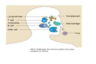
Classical pathway. Alternative pathway. Lectin pathway. Antigen:antibody. Bacterial surface. MBP: Bacterial surface. The complement. Complement activation. Recruitment of inflammatory cells. Opsonisation. Killing of pathogen. C3. DAF. B. Tick over conversion. Bb. C3.

E N D
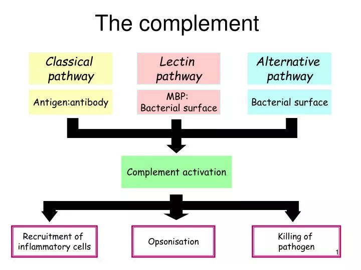
Classical pathway Alternative pathway Lectin pathway Antigen:antibody Bacterial surface MBP: Bacterial surface The complement Complement activation Recruitment of inflammatory cells Opsonisation Killing of pathogen
C3 DAF B Tick over conversion Bb C3 C3b C3b Binding to the surface of a microorganism stabilises C3b Ba D Classical pathway antigen antibody C2b C4 C2 + The classical pathway of complement activation is actually a mechanism of enhancing the adaptive immune response. In contrast, the "alternative" pathway of complement activation does not require antibody for triggering of the cascade. C1r C1s C1q C4a C4b C4b2a Decay Accelerating Factor C3a C3b Alternative pathway
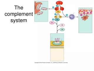
C3 C3 Complement links innate and adaptive immunity • Components of the complement cascade can bind to the surface of invading organisms. This bound complement can then:- • Trigger the complement cascade via the alternative pathway, ultimately leading to assembly of the membrane attack complex and the destruction of the organism. • Enable phagocytosis of the organism by cells bearing complement receptors. Microorganisms Alternative pathway (innate immunity) Inflammation C5a chemotaxis anaphylotoxin lysis MB-lectin pathway C6 C5b C7 C9 C3a C3a C8 membrane membrane attack complex cytoplasm C3b C3b Antibody/antigen complexes complement receptors phagocytosis Classical pathway (adaptive immunity)
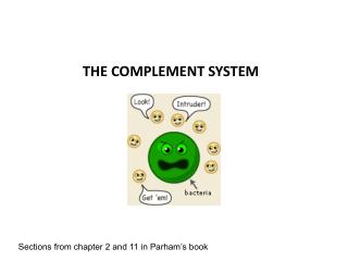
There are three ways to trigger the complement cascade Lectin binding to pathogen surfaces Microorganism surfaces Antibody/antigen complexes Alternative pathway MB-lectin pathway Classical pathway C3 convertase C3a C3 C3b C3b C5b, C6, C7, C8 and C9 C3b C3a and C5a Membrane attack complex Complement receptors on phagocytic cells Inflammation Chemotaxis Opsonisation N.B. Brittany Spaniels is an example for C3 deficiency Immune complex formation
Where do antibodies come from? Antibodies are produced by B cells, and in mammals B cells originate in the foetal liver and bone marrow. The “B” in B-cell refers to “bursa-derived”. In birds, a specialized organ known as the bursa of Fabricius (the organ was first described in the notes of Hieronymus Fabricius published posthumously in 1621). A dorsal diverticulum of the cloaca, the bursa is responsible for antibody production in newly hatched chicks. The bursa of Fabricius is absent in mammals, B cell development occurring in the bone marrow. A notable exception is the rabbit, where the sacculus rotundus (located at the ileo-coecal valve) performs a similar role.
Antibody Complement fixation Antigen binding Fc receptor binding Myelomas are clonal cancers of plasma cells. The terms “antibody” and “immunoglobulin” are synonyms.
The basic structure of immunoglobulin Variable domain CL VL1 Light chain Heavy chain Fab CH1 VH1 CH2 Hinge region Fc CH3 pepsin C papain C N N
Antibody variation Idiotypic variation - variation in the antigen binding domain V VH D V J J VL CH1 CL Allotypic variation - variation in the amino acid sequence of the heavy and light chain genes (inherited) CH2 Isotypic variation - is the heavy chain g,m,e,a or d and the light chain k or l? CH3 CH4
conformational epitope antibody linear epitope Antigen polypeptide Antibody Antigen binding site epitope polypeptide protein antigen Types of epitopes Epitopes can be “linear” or “conformational” Bind “antigen” “lock and key” The distinction between conformational and linear epitopes is important in the design of vaccines and immunological assays based on peptides Anti-serum”
Antibody plays a central role in the response to an invading pathogen Antibody bound to the surface of an invading organism can:- • Trigger antibody-dependent cell-mediated cytotoxicity "ADCC" virus infected cell • Fix complement enabling phagocytosis of the organism by cells expressing complement receptors and triggering of the "classical" pathway of the complement cascade • Enable phagocytosis of the organism by cells expressing Fc receptors
IgG • Secondary response to antigen • Fc receptor binding • Neutralises antigen, fixes complement • Tends to be high affinity due to affinity maturation Fab Fc IgG3 In humans there are subclasses of IgG IgG1, IgG2, IgG3, IgG4 Each class has a different function
The primary and secondary antibody response Primary exposure Secondary exposure to antigen to antigen Primary Secondary response response 100,000 IgG 10,000 1000 Log antibody titre 100 10 IgM 1 0 0 7 14 21 28 35 42 Days • The primary response is delayed, IgM appears first followed by IgG. • The secondary response is rapid, IgG appears first and achieves a higher titre than in the primary response. • Passive immunity is conferred to new-born animals by colostral transfer.
IgM Pentameric structure Primary response to antigen Often low affinity but high avidity Neutralises antigen, fixes complement Low affinity receptor on monocytes (FcmR) IgM J - chain bacterium
Heavy glycosylation of hinge region J-chain Secretory component IgA Dimeric or tetrameric structure Secreted immunoglobulin, resistant to proteolytic degradation Secretory component mediates transport across epithelial surfaces High affinity receptor on monocytes and neutrophils (FcaR) In humans 40mg/kg body weight of polymeric IgA (pIgA) is translocated to the gut every day; the total daily production of IgG is estimated at 30mg/kg.
IgE Triggers the release of histamine, mast cell degranulation High affinity receptor for IgE is expressed on mast cells and basophils (FcεRI), lower affinity receptor on monocytes (FcεRII)
VH VL Cg1 CL Cg2 Cg3 IgG IgY VH VH VH VL VL VL Ce1 Cu1 Cu1 CL CL CL Cu2 Cu2 Ce2 Cu3 Ce3 IgY(DFc) hinge region Cu4 Ce4 IgY IgE IgM is thought to be the ancestor to all Ig classes (it is universally distributed in vertebrates). However, while the dominant low molecular weight serum antibody in mammals is IgG, birds reptiles, amphibia and lungfish have IgY. IgY is the ancestor of both IgG and IgE. Anseriform birds express IgYDFc, a truncated IgY molecule.
VH VH VL VL Cg1 Cg1 CL CL Cg2 Cg2 Cg3 Cg3 …and then there is the camelids. VH VH Cg2 Cg2 Cg3 Cg3 Four-chain camel IgG1 Long-hinge heavy chain camel IgG (IgG2) Short-hinge heavy chain camel IgG (IgG3) Human IgG