Download
1 / 1
10 likes | 115 Views
Survey Specifics of Nucleus Pulposus Cells of Human Intervertebral Disc in Chitosan-Gelatin and Alginate Scaffolds Masoud Ghorbani 1 * , Hamid Bahramian Renani 2 , Batool Hashemibeni Beni 2 , Zeinab Karimi 2

E N D
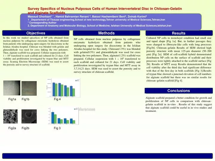
Survey Specifics of Nucleus Pulposus Cells of Human Intervertebral Disc in Chitosan-Gelatin and Alginate Scaffolds • MasoudGhorbani1* , Hamid BahramianRenani2, BatoolHashemibeni Beni2, Zeinab Karimi2 • 1. Departement of Tissuseengineering,School of new technology,Tehran university of Medical Sciences,Tehran,Iran • * Coressponding Author • 2. Department of Anatomy and Molecular Biology, School of Medicine, Isfahan University of Medical Sciences,Isfahan,Iran • * Objectives Methods Results In this work we studied specefices of NP cells obtained from nucleus pulposus by collagenase enzymatic hydrolysis obtained from patients who undergoing open surgery for discectomy in the Isfahan Alzahra hospital. Chitosan was blended with gelatin and glutaraldehyde was used for cross linking the two polymers. Then, alginate scaffold was prepared. Cellular suspension with 1 × 10⁵ transfered to each scaffold and cultured for 21 days. Cell viability and proliferation investigated by trypan blue and MTT assay. Scaning Electron Microscopy (SEM) was used to assert the porosity and to survey structure of scaffold. Cultured NP cells in monolayer condition had small size and taped shape [Fig 1a]. But, in further passages they were changed to fibrocyte-like cells with long processes [Fig1b]. Chitosan–gelatin Results of SEM showed high porosity structure with mean 125-μm diameter (50–200 μm) [Fig 2a]. SEM of cell-scaffold hybrid demonstrated distribution NP cells on the surface of scaffold and their processes were tightly attached to the scaffold surface [Fig 2b]. Results of MTT assay Results demonstrated that the cell viability after the third day had significant difference with that of the first day in both scaffolds [Fig 3].Results of trypan blue showed a punctual elevation of cell numbers for alginate scaffold but there was no similar results for chitosan–gelatin scaffold [Fig 4]. NP cells obtained from nucleus pulposus by collagenase enzymatic hydrolysis obtained from patients who undergoing open surgery for discectomy in the Isfahan Alzahra hospital for this study. Chitosan(1.5%) was blended with gelatin(0.5%) and glutaraldehyde was used for cross linking the two polymers. Then, alginate(1.2%) scaffold was prepared. Cellular suspension with 1 × 10⁵ transferred to each scaffold and cultured for 21 days. Cell viability and proliferation investigated by trypan blue and MTT assay in 3,7,14,21 days. SEM was used to assert the porosity and to survey structure of chitosan scaffold. Conclusions Ffiیبیبییبdjkjk پییReferences Alginate scaffold prepared a better condition for growth and proliferation of NP cells in comparison with chitosan–gelatin scaffold in in-vitro . Results of this study suggest that alginate scaffold could be useful in in vivo studies and treatment.