Download

1 / 21
210 likes | 368 Views
Regulation of Cell Division. M. anaphase. metaphase. telophase. prophase. C. G 2. interphase (G 1 , S, G 2 phases). mitosis (M). cytokinesis (C). G 1. S. Frequency of cell division. Frequency of cell division varies by cell type embryo cell cycle < 20 minute skin cells

E N D
M anaphase metaphase telophase prophase C G2 interphase (G1, S, G2 phases) mitosis (M) cytokinesis (C) G1 S Frequency of cell division • Frequency of cell division varies by cell type • embryo • cell cycle < 20 minute • skin cells • divide frequently throughout life • 12-24 hours cycle • liver cells • retain ability to divide, but keep it in reserve • divide once every year or two • mature nerve cells & muscle cells • do not divide at all after maturity • permanently in G0
sister chromatids centromere single-stranded chromosomes double-stranded chromosomes There’s noturning back, now! Overview of Cell Cycle Control • Two irreversible points in cell cycle • replication of genetic material • separation of sister chromatids • Checkpoints • process is assessed & possibly halted
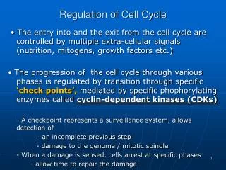
Checkpoint control system • Checkpoints • cell cycle controlled by STOP & GO chemical signals at critical points • signals indicate if key cellular processes have been completed correctly
Checkpoint control system • 3 major checkpoints: • G1/S • can DNA synthesis begin? • G2/M • has DNA synthesis been completed correctly? • commitment to mitosis • spindle checkpoint • are all chromosomes attached to spindle? • can sister chromatids separate correctly?
G1/S checkpoint • G1/S checkpoint is most critical • primary decision point • “restriction point” • if cell receives “GO” signal, it divides • internal signals: cell growth (size), cell nutrition • external signals: “growth factors” • if cell does not receive signal, it exits cycle & switches to G0 phase • non-dividing, working state
How this works Primary mechanism of control • phosphorylation • Cyclin dependent kinase are also present in the cell in the inactive form • When growth factors cause cyclin to be produced it activats cyclin dependent kinase (CDK) • CDK that can activate proteins needed to push the cell into mitosis
inactivated Cdk Cell cycle signals • Cell cycle controls • cyclins • regulatory proteins • levels cycle in the cell • Cdks • cyclin-dependent kinases • phosphorylates cellular proteins • activates or inactivates proteins • Cdk-cyclin complex • triggers passage through different stages of cell cycle activated Cdk
Leland H. Hartwell checkpoints Tim Hunt Cdks Sir Paul Nurse cyclins 1970s-80s | 2001 Cyclins & Cdks • This happens a lot of the checkpoints. Interaction of Cdk’s & different cyclins triggers the stages of the cell cycle
Spindle checkpoint G2 / M checkpoint Chromosomes attached at metaphase plate • Replication completed • DNA integrity Inactive Active Active Inactive Cdk / G2cyclin (MPF) M cytokinesis APC C mitosis G2 G1 S Cdk / G1cyclin Inactive MPF = Mitosis Promoting Factor APC = Anaphase Promoting Complex Active G1 / S checkpoint • Growth factors • Nutritional state of cell • Size of cell
APC CHECKPOINT • The spindle checkpoint inhibits the APC until all sister-kinetochores are attached to opposite poles of the mitotic spindle. • When all kinetochores are properly attached the spindle checkpoint is silenced and the APC becomes active. • The activated APC then targets securin for degradation. Securin inhibits a protease called separase, which cleaves cohesins allowing anaphase onset.
External signals • Growth factors • coordination between cells • protein signals released by body cells that stimulate other cells to divide • density-dependent inhibition • crowded cells stop dividing • each cell binds a bit of growth factor • not enough activator left to trigger division in any one cell • anchorage dependence • to divide cells must be attached to a substrate • “touch sensor” receptors
Growth Factors and Cancer • Growth factors can create cancers • proto-oncogenes • normally activates cell division • growth factor genes • become oncogenes (cancer-causing) when mutated • if switched “ON” can cause cancer • example: RAS (activates cyclins) • tumor-suppressor genes • normally inhibits cell division • if switched “OFF” can cause cancer • example: p53
Cancer & Cell Growth • Cancer is essentially a failure of cell division control • unrestrained, uncontrolled cell growth • What control is lost? • lose checkpoint stops • gene p53 plays a key role in G1/S restriction point • p53 protein halts cell division if it detects damaged DNA • options: • stimulates repair enzymes to fix DNA • forces cell into G0 resting stage • keeps cell in G1 arrest • causes apoptosis of damaged cell • ALL cancers have to shut down p53 activity p53 is theCell CycleEnforcer p53 discovered at Stony Brook by Dr. Arnold Levine
p53 — master regulator gene NORMAL p53 p53 allows cells with repaired DNA to divide. p53 protein DNA repair enzyme p53 protein Step 2 Step 1 Step 3 DNA damage is caused by heat, radiation, or chemicals. Cell division stops, and p53 triggers enzymes to repair damaged region. p53 triggers the destruction of cells damaged beyond repair. ABNORMAL p53 abnormal p53 protein cancer cell Step 2 Step 1 Step 3 The p53 protein fails to stop cell division and repair DNA. Cell divides without repair to damaged DNA. DNA damage is caused by heat, radiation, or chemicals. Damaged cells continue to divide. If other damage accumulates, the cell can turn cancerous.
Development of Cancer • Cancer develops only after a cell experiences ~6 key mutations (“hits”) • unlimited growth • turn on growth promoter genes • ignore checkpoints • turn off tumor suppressor genes (p53) • escape apoptosis • turn off suicide genes • immortality = unlimited divisions • turn on chromosome maintenance genes • promotes blood vessel growth • turn on blood vessel growth genes • overcome anchor & density dependence • turn off touch-sensor gene It’s like anout-of-controlcar with manysystems failing!
What causes these “hits”? • Mutations in cells can be triggered by • UV radiation • chemical exposure • radiation exposure • heat • cigarette smoke • pollution • age • genetics
Tumors • Mass of abnormal cells • Benign tumor • abnormal cells remain at original site as a lump • p53 has halted cell divisions • most do not cause serious problems &can be removed by surgery • Malignant tumor • cells leave original site • lose attachment to nearby cells • carried by blood & lymph system to other tissues • start more tumors =metastasis • impair functions of organs throughout body
Traditional treatments for cancers • Treatments target rapidly dividing cells • high-energy radiation • kills rapidly dividing cells • chemotherapy • stop DNA replication • stop mitosis & cytokinesis • stop blood vessel growth
New “miracle drugs” • Drugs targeting proteins (enzymes) found only in cancer cells • Gleevec • treatment for adult leukemia (CML)& stomach cancer (GIST) • 1st successful drug targeting only cancer cells withoutGleevec withGleevec Novartes