Download
1 / 33
330 likes | 483 Views
Granulomatosis Colitis. Presented by Dr. Leon Wolf. History . C.C. Anemia and HO + 45 yo male asymptomatic PMH h/o goiter, Rx Synthroid FH CAD DM Colonic polyps SH born outside of USA, postal worker ROS w/o wt loss, fever w/o cough, sputum hemoptysis. Physical Exam.

E N D
Granulomatosis Colitis Presented by Dr. Leon Wolf
History • C.C. Anemia and HO + • 45 yo male asymptomatic • PMH h/o goiter, Rx Synthroid • FH CAD DM Colonic polyps • SH born outside of USA, postal worker • ROS w/o wt loss, fever w/o cough, sputum hemoptysis
Physical Exam • Healthy appearing wt.220 T.98.6 • HEENT R. neck fullness • Lungs clear • Abd soft w/o masses, LSKK • Rectal w/o masses, HO+ • Ext w/o joint fullness or tenderness • Skin w/o rashes
LAB • Hgb 10.6, MCV 77 • WBC 8,900 ; normal differential • CMP normal • CEA 1.4
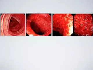
ENDOSCOPIC EVALUATION • Colon cecal villous,nodular friable lesion • EGD gastric erythema esophageal nodule • Microscopic Colon: granulomatous colitis Stomach: mild gastritis Esophagus: papilloma
Clinical Course • RX Pentasa, iron • CXR negative • SBFT negative • CTABD/PELVIS negative • PPD positive 20yrs ago
Re-Colonoscopy • Villous, nodular lesion • Open ileocecal valve • Ileal lymphoid hyperplasia • Cultures AFB,Fungus, O&P • Stains
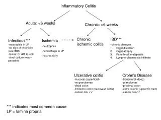
Gastrointestinal diseases Inflammatory bowel disease Crohn’s disease Ulcerative colitis Nodular lymphoid hyperplasia Celiac disease Necrotizing enterocolitis Gastrointestinal diseases continued Behçet’s disease Eosinophilic gastroenteritis Hirschsprung’s disease with necrotizing enterocolitis Neoplasms Anatomical or vascular abnormalities Diseases to Consider in the Differential Diagnosis
Hematologic diseases Chronic granulomatous disease Langerhans’ –cell histiocytosis Familial hemophagocytic lymphohistiocytosis Systemic inflammatory diseases Sarcoidosis Wegener’s granulomatosis Juvenile dermatomyositis Juvenile rheumatoid arthritis Systemic lupus erythematosus Diseases to Consider in the Differential Diagnosis Continued
Diseases to Consider in the Differential Diagnosis continued • Infectious diseases • Mycobacterium tuberculosis infection • M. avium infection • Yersinia infection • Giardia lamblia infection • Tropheryma whippelii infection • Bartonella henselae infection
Differential DX • Yersinia • Sarcoidosis • Crohn’s disease • Tuberculosis
Yersinia • Gram negative rod • Contaminated milk, milk products • Acute manifestations • Enterocolits most common <5 yo • Adenitis, ileitis >5 yo • Bacteremia in pts underlying disease • Reiter’s syndrome • Self limited 3 to 4 wks
Sarcoidosis • Gastrointestinal involvement uncommon other than liver granulomatosis • Stomach primarily,bleeding ulcerations • Small intestine nodal or lymphatic blockage • Esophageal obstruction lymph nodes or infiltration • Pulmonary or renal involvement with above
Tuberculosis • Koch 1882 ID bacillus • Primary pulmonary disease • Pre antiboitics 55-90% GI involvement • Proportional to pulmonary disease • Post antiboitics GI disease have <50% pulmonary tb evidence
Tuberculosis organisms • M. tuberculosis • M.bovis • (M. avium)
Patients At Higher Risk • Immigrants (travel endemic areas) • AIDS • Urban poor • Living on reservations • Prisoners • NH residents
Gastrointestinal Areas • Ileocecal/ileal approx 75% • Asc.colon appendix approx 20% • Uncommon jejunum,stomach,esophagus, sigmoid/rectum, anal • Multiple areas-skip areas
Clinical Sx and Exam • Non-specific sx 80-90% pain wt loss diarrhea/constipation blood in stools • PE abdominal mass perianal lesions
Complications • Hemorrhage • Perforation • Obstruction • Fistula formation • Malabsorption
Endoscopic Findings • Ulcerative 60% • Hypertrophic 10% • Mixed 30% • Circumferential ulcers • Scarred open IC valve
Radiological Findings • BE/SBFT ulcers thickening/distortion stenosis pseudopolyps • CT adenopathy-central necrosis mass calcified nodes
Diagnosis • Stain <20% • PCR 80% • Culture <30% mucosal biopsies ? % surgical specimen esp node n.g. stool esp with pulm disease • Presumptive +PPD, +CXR • Therapeutic Response
Clinical Course • Iron RX increase hgb felt less dizzy • + AFB culture • M.gordonia
Ten Diseases Doctors Miss Reader’s Digest Feb 2003 • Hepatits C • Lupus • Celiac Disease • Hemochromatosis • Aneurysm • Lyme Disease • Hypothyroidism • Polycystic Ovary Syndrome • Chlamydia • Sleep Apnea