Download

1 / 46

E N D
case • A 34 years old man presented to emergency with loss of consciousness at his work and brought by his friend to hospital who mentioned that all of sudden while he is sitting , He collapsed on the ground with spasm all over and abnormal sound ;the symptoms subsided within 2-3 minutes and his cons. recovered after 60 minutes , after that; He was oriented with sever headache and minimal deficit on right side, return back his normal life in next few hours. • Neurological exam: normal with mild right side weakness affecting UL and UL+upper facial weakness • What is next step???? Main problem? D D
D D • TIA • Seizure • Migraine • Syncope • Psychogenic
SEZURE? What is seizure • PARTIAL OR GENERALISED? • Is it epileptic? • Early or late epilepsy and why? • Idiopathic , or symptomatic or cryptogenic? • Primary G. or secondary with focal elements? • Next on arrival ? Treatment or D. • Treatment line?
Epilepsy and seizure disorders Hayder k. Hassoun prof. Of clinical neurology Kufa college of medicine-department of neurology
Seizure? Latin ward sacire , to take possession off • It is paroxysmal event due to abnormal excessive hyper synchronous central neuronal discharge, with various clinical manifestation ranging from dramatic convulsion activity to phenomenon may not be seen by an observer depending on extent and area affected .what is convulsion? • What is epilepsy? Recurrent unprovoked seizures due underlying unproven structural or functional disorder . • The following are not consider as epilepsy we call it symptomatic seizure (provoked seizure)= not persist if underlying disorder is corrected. • Seizure in course of medical and N . illness with temporary deranged brain function. • Seizure due to reaction of brain to PSL stress from sleep deprivation, fever, sedative and alcohol withdrawal (low seizure threshold) • Isolated seizure without recognized cause
When seizure occurs without any identifiable cause it is called cryptogenic seizure (probable symptomatic). • When seizure occurs with genetic mediation it is called idiopathic seizure. • When both associated with recurrent unprovoked attacks more than 2 = epilepsy. According to age of onset: • Early onset E.=E. below age of 25 years.? • Late onset E. =E. after age of 25 years.?
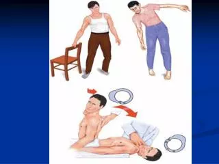
Seizure versus syncope & psychogenic • Episodic recoverable loss of consciousness other than asleep are due to –either faint or fit (seizure). • more commonly is due to inadequate C. Blood supply ( syncope). • Seizure loss of consc. Is due to global C. dysfunction due to abnormal C. Discharge. Differentiation mainly by history. • Syncope is brief feeling of light headedness often precedes faint then V. Darkens ringing in ear often provoked by emotional Factors, and in stand position except if it is due to cardiac cause which can happen in stand ,exercise or flat position.
Incidence and etiology: • *0,2-0,5% epilepsy incidence in various pop. Prevalence is 5-10/1000. • *5-10% of pop. Will have at least one seizure in their life with higher incidence occur in childhood and adulthood. • *male :female is 1.5:1 . • Causes of seizure and epilepsy • Seizures are a result of a shift in the normal balance of excitation and inhibition in CNS. • There are many factors which might disturb this balance resulting in seizures? Our understanding of mechanisms involved is very limited, however the dynamic interplay of the following factors is likely to cause seizure or epilepsy in a given pt.
1-The normal brain is capable of having seizure at certain circumstances and there are differences between individuals in susceptibility or threshold for s. .ex: febrile convulsion which is induced by high fever 3-5% of children but not all? • low s. threshold child? • Endogenous --------clearly genetic • Developmental----- s. threshold (balance) is vary from person to another developmentally and it is different at different maturation stages 2-Variety of condition have extremely high likelihood of resulting in chronic s. disorder (epileptogenic) ex: sever penetrating head trauma carry 50% risk of having epilepsy. This process of transforming normal neural network in an abnormally hyper excitable called epileptogenesis and specific factor responsible called epileptogenic factor .these include trauma ,infection, tumor, stroke------including genetic factors like in JME. And benign familial neonatal C. .
3-seizures are episodic that it is to say we have many patient free of s. for months and Years between their s. ? precipitating factors that induce s. in epileptic person ,these are • intrinsic PSL factors as sleep deprivation ,psychological or physical stress and hormonal changes associated with m. cycle • extrinsic factors such as exposure to toxic substance and drugs • Genetic factors? growing no. of E. are recognized to be caused by specific gene defect while many other have clear hereditary influence but clear gene defect are not recognizes (common). At least 3 of genetic epilepsy are known to be channelopathies. • Neuronal migration defect is readily recognized by MRI and it is important cause of acquired and genetic epilepsy.
conclusion : the role of each factor mentioned needs to be carefully identified for appropriate management • Genetic predisposing factor (family h. of e.) in pt. with FC is indication for closer follow up and more diagnostic evaluations.
Causes of symptomatic (secondary) seizure • Acute electrolyte disturbance: Hyponatremia <120 mEq/L--- especially acute. Acute hypernatremia >155 mEq/L--- especially acute Hyperosmolarity >310 mOsm/l Hypocalcemia< 7mg/dl Hypoglycemia <30mg/dl • Drugs and toxins Isoniazid, penicillin, theophylline, aminophylline , ephedrine, phenylpropanolamine, terbutaline, lidocaine ,meperidine TCA Cyclosporine , Cocaine,phencyclidine, amphetamine ,alcohol withdrawal. • Hepatic encephalopathy , • renal failure • Central nervous system Hypertensive encephalopathy, eclampsia Sickle cell anemia , thrombotic thrombocytopenia purpura SLE Meningitis, encephalitis ,brain abscess Acute head trauma (impact) ,stroke, brain tumor
classification • it is essential step in the evaluation and management. • ILAE 1981 published a classification still applied with some • modification. they depend on clinical feature and associated EEG finding. • depending on a concept whether most or only part of cerebral • cortex is involved in the beginning as well area of origin and subsequent pattern of spread in brain ex; focal (partial motor E.) ,so the s. is of 2 types • partial (focal) • generalized. The later the s. involve diffuse region of brain simultaneously and bilaterally symmetrical. this may result from cellular ,biochemical, or structural abnormality of brain in contrast to focal s. which is typically associated with underlying structural abnormality of brain.
partial seizure (simple or complex): indicate focal, localized area of discharge. • 40% of children s. and 70% of adult s. are partial. • Simple partial (SP):when the discharge is remained localized to focal area of cortex ,clinically the pt . is reacting normally to environment. It may be considered as an auras to complex partial or G. seizure. Specification of symptoms are of great help in localization of discharging focus? locus can be • SP motor S. (frontal lobe)? when s. is with motor signs as clonic (rhythmic jerking) or tonic (stiffening) movement usually involving face or and hand .it may spreads to involve the adjacent areas ex; ie, start in thumb to finger to face to leg this is called jacksonian march seizure . • arm extension • speech arrest or aphasia. SP sensory S : as sensory symptoms .it is originating from parietal lobe SP temporal S: foul odors (uncus amygdale), visual changes (middle &inf. Temporal area), dejavu &jamas vu (hipocampus and parahipocampus area), fear ,pleasure, anger dreamy sensation and forced thinking (parahipocampus-septal area) or voice or music (auditory ass. area) or as lip smacking ,abdominal symptoms and cardiac arrhythmias SP occipital S. : visual s. as metamorphopsias. • SPS may followed by Todd’s palsy .it is transient focal neurological abnormality last for 24-48 hours which is secondary to post ictal depression of epileptogenic cortical areas ex: focal weakness follow SPMS, numbness follow SPSS . • THESE reflect the focal nature of seizure and it is of localizing value to site of seizure.
about 70% of adult and 40% of children with new onset epilepsy have partial (focal) seizure. • in many cases it is not possible to a specific cause. although focal seizure imply a cerebral injury or lesion. the most specific and common causes are: • hipocampal sclerosis ,gangliogliomas ,Glial tumor, cavernous malformation ,cortical dysplasia or agenesis (neuronal migration defect) and hamartomas. Encephalitis ,hemorrhage and trauma. • Not all c. lesion produce E. but how particular lesion become epileptogenic ? poorly understood.*
Complex Partial S. :CPS • There is impaired sensorial =unresponsiveness=dissociation? • 80% of cases the focus in temporal lobe and 2/3 is from Mesial TL structure. • 20% , the focus from frontal lobe. • Many are evolving from SPS and may progress into G. seizure. when the discharge is spreads bilaterally involving both cc. • It is usually start as SPS as an aura ex: bad odors (uncinate fit) or ant temporal lobe or limbic symptoms then progress within seconds to CPS as the sensorium impaired with or without automatism. • Psychomotor ,temporal and limbic s. are not synonyms as previously have been described ? but they are used to describe ictal behavior now classified as CPS.----
Generalized seizure: • discharge is involving both H. from the onset (loss of consciousness) with lacking of clinical and EEG feature that suggest localized c. discharge. • it should be differentiated from focal s. that spread to cause G. seizure in this case we call it secondary G. clinically the G seizure classified into • grand mal (tonic clonic convulsion): • start as loss of c. –falling –tonic phase with epileptic cry then clonic with or without incontinence or tongue bite ,all fit usually not last more than 90 sec. followed by post ictal phase which manifested as transient deep stupor followed in 15-30 min by lethargy ,confused state with automatic behavior ,as recovery progress many pt have headache ,muscle snoring ,lack of energy and mood change may last for24hr . • PSL changes in GS? • complication? • can sudden death occur ?SUDEP
Absence seizure (petit mal) : typical one occur mainly in children characterized as brief momentary lapse in awareness, staring, rhythmic blinking and some times clonic jerks in arms and face .no postictal phase and no recalling to events .each absence last for 10 sec. (EEG is 3 spike and wave /sec), good response to AED. • Genetic factors play a role .some patient may develop grand mal s. in future • Atypical absence: more gradual ,not resolve abruptly and accompanied by autonomic features and loss of postural tone (drop). it is seen mainly in children with MR and not respond well to AED. • Myoclonic s.? • Atonic s.?
B A A: Stage 1; Freezing, Sniffing B: Stage 2; Head nodding
D C C: Stage 3; Contralateral forelimb clonus D: Stage 4; Forelimb Clonus and rearing
E E E: Stage 5; Rearing and Falling Fully kindled rate (chemical or LFS)
Some of clinical types? • Febrile convulsion • -Most common convulsion in children. it affect 3%-5% of children • -age: 6 months-5 years ,may be happened at age of 6,7 some times • -30% have more than one attack, recurrence is greatest when there is positive family H. or when 1st seizure occur <1 year. • -it increase the like hood of E. ,but risk is not more than 3-5%, reaching to 10-13% in those have FH of E. ,prolonged or focal S. , or who have abnormal N. examination. • -it not increase or cause MR, poor school performance or behavior changes. • Benign partial epilepsy of childhood with central-midtemporal spikes (rolandic epilepsy)? • -it form about 15% of all pediatric E. . It is common between 4-13 years • -normal children ,mainly during sleep noticed as generalized while it is focal to start with. if it occurs during day time are typically focal involve twitching one side of face ,paraesthesia.it ,drooling ,or speech arrest, may progress to cause hemitonic posture or clonic movement. • -EEG is characteristic. it disappear at adolescence. • outcome not affected by treatment.
Juvenile myoclonic epilepsy? 8-20 years of age ,positive FH, it goes with AD inheritance • Lenox-gastaut syndrome? • Heterogeneous group of early childhood epileptic encephalopathies ,seen in an abnormal brain str. function ,MR, with uncontrolled S. • Temporal lobe epilepsy? • -MOST common E. syndrome of adult,40% of adult E. , start in late childhood or adolescence and often there is H .of febrile C. • -virtually all pt have complex partial S. • -it ‘s origin from mesial temporal limbic str. typically with characteristic lesion known as hipocampal sclerosis. • -20% is caused by other str lesion including cavernous malformation, hamartomas ,cortical dysplasia , Glial tumor and scars related to previous trauma or encephalitis.
Post traumatic epilepsy? • -it related directly to severity of HT. especially within 1-2 years ,occasionally even after 5 years or more. • -1/3 of sever head injured pt . have epilepsy within 1st year. • Sever head injury defined as HT with : cerebral contusion ,intracerebral or IC bleeding, unconsciousness or prolonged amnesia >24 hrs , persistent N. abnormality. • -2/3 have partial S. or secondary G. • -mild HT: (uncomplicated brief loss of C. ,no skull fracture ,no contusion or hematoma and no focal N. signs) don’t increase risk of S. .
Investigation? • EEG- IS to confirm and support the diagnosis, classify S ,identifying E syndrome and for therapeutic decision. • Neuroimaging: • MRI: complement to EEG and can detect str. brain pathology that may be causally related to development of E. as hipocampal sclerosis .hamartomas , cortical dysplasia and cavernous malformation. • Indication? • What about PET AND SPECT? offer functional view of brain by using PSL active radio labeled tracers to image brain’s metabolic activity PET & BF as SPECT. • other test: blood tests , CSF , ECG • DD: other non epileptic episodic disorder that may mimic s. • Movement disorder • Migraine • Behavior and psychiatric : as pseudo -seizure • Cataplexy • Syncope and convulsive syncope • TIA • Alcohol blackout and hypoglycemia
DIAGNOSIS? and management • -determine if pt have E . or not---------??????? • -classify it . • -try to identify if possible underlying cause . • History? 1st attack or he is known case , Age (onset) is important. • Examination? • Investigation?
Treatment: *AED is unnecessary in cases of symptomatic S. when the cause is corrected or in cases of single unprovoked S. with normal clinical and lab . finding where the recurrence is not happened. • *Those with recurrent S or even single with possible high recurrence (focal N . finding on clinical or radiological or EEG exam.) • *the goal of treatment to stop the attack completely, taking the following in consideration: • Select AED for particular S. type? • If S . persist upon reaching toxic AED level or major SE occurs the you have to choose the alternative • Do not stop the 1st one unless you introduce the another. • If the S . continue in spite of 2 medication ,consider referral to specialized center for further monitoring ,complex combination therapy and possible surgical treatment. • Toxic level of some AED may cause S. (like phenytoin and Carbamazepine)
AED in relation to S type: • Simple and complex P. S.& Sec. G. : • carbamazapine, phenytoin, gabapentin, lamotrigine and Topiramate • Primary G : • tonic clonic----------valproate ,carbamazapine. ,phenytoin , lamotrigine ? • absence--------------ethosuximide ,valproate ,lamotrigine? • myoclonic and tonic-------valproate ,clonazepam, Levitrecetam, zonisamide , Topiramate.
STATUS E. • This can be of G or partial type .the generalized motor status is important one as it carry risk of permanent brain damage and death. • It is repeated S. for 5-10 min. from which the pt. not regain cons. In between. • Common causes? • Treatment is urgent? • 1-within 1st 5 minutes: • Give O2, ensure ABC. Monitor vital signs ,ECG , and oximetry • Establish iv access and send blood sample for glucose, electrolyte, drug level , CBC and toxins. • Give glucose (with thiamine in adult)? • AED immediately? • 1st line: depend on availability, IV lorazepam 0.1mg/kg in rate of 2mg/min Ordiazepam .2mg/kg in rate of 5mg/min with phenytoin (phenytoin 18-20mg/kg in a rate of 50mg/min in adult and 1mg/kg/min in children). (Phosphenytoin is alternative to phenytoin), • If S. Persist you can repeat diazepam dose after 5 min. ,or you can use 2nd line treatment simultaneously to maintain recovery or alternative to above as drugs Depakine like valproate 20-40mg/kg infusion up to 3mg/kg/min safe up to 6mg/kg/min or occasionally Phenobarbital when the patient at special care unit. • Or IM midazolam up to 10 mg IM can be used or valium rectally if no IV access is available specially as pre hospital treatment.
2-Within 30 minutes: (refractory status) =No response to above lines • Admission to RCU intubation may be required with short acting paralytic ex; succinylecholine for midazolam loading (.1mg /kg up to 4 mg IV at 2mg/min) or Phenobarbital 5mg/kg IV at 50/min, the pt. should be in RCU with availability of V. Assistance and EEG monitoring. • Simultaneously phenytoin, Phosphenytoin or Depakine (2nd line drugs) should be used as mentioned above to maintain recovery. • If patient responding maintenance treatment should be started. • Last step GA may need (propfol or thiopental) EEG monitoring is important and NM blockade agent may need.
Epilepsy surgery • Indicated only in cases of intractable S. (uncontrolled in spite of adequate trial of 2 appropriate single agent or combination therapy of 2 agent ) • Incidence of IE : 20% with considerable risk of S. complication ,inability to drive ,stigmatization by school, employers and families. • How surgery is of help? • Psychosocial problems? Related to type and control of E. • Pregnancy and E. ? • -90% they will have uneventful P. . • -in 4-10% of offspring will have kind of S. ,in comparism to 1.5% in GP. • -1.5-3 fold increase rate of P. complication? As premature labor, placenta abruption ,bleeding ,toxemia ,still birth and AED complication----- • -AED? • -Safe Drug? Tegretol , Lamotrigine for partial and generalized And Ethosuximide (for absence) • -Treatment? It is high risk P.?
side effect of AED Dose related Idiosyncratic
In children with a single seizure, the rate of recurrence was 27% when routine EEG was normal, 37% with a nonepileptiform EEG abnormality, and 58% when the EEG showed epileptiform activity.1 In adults with a single idiopathic seizure and EEG with epileptiform discharges, the 2-year recurrence rate is 83%, with an observed sensitivity of 48% and specificity of 91% for EEG predicting recurrence. In a meta-analysis, the combination of remote symptomatic etiology and abnormal EEG predicted a 65% risk of future seizure. For these patients, it may be appropriate to consider treating with an AED. In contrast, for a single idiopathic seizure with a normal EEG, the 2-year recurrence risk following first seizure is around 30 to 40%.Unless special circumstances exist, medication is generally not started in this situation.
Factors Predicting a HigherRecurrence Risk Following a Single Unprovoked Seizure • Abnormal neurologic examination • Abnormal electroencephalogram • Abnormal head imaging • Partial seizure • Status epilepticus • Remote symptomatic etiology • Family history of epilepsy