Download
1 / 53
540 likes | 687 Views
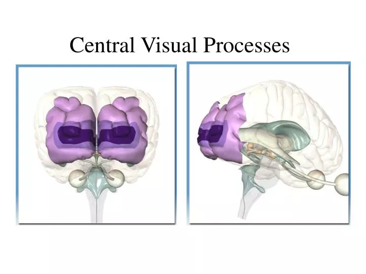
Central Visual Processes. Central Visual Pathways. Primary Visual Cortex Receptive Field Columns Hypercolumns Spatial Frequency Nerve or Cortical Damage Higher Visual Areas. Occipital Lobe. Occipital Lobe: Calcarine Sulcus -- V1 -- Striate Cortex. Cells in V1.

E N D
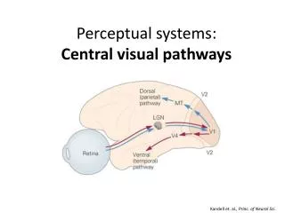
Central Visual Pathways • Primary Visual CortexReceptive Field • Columns • Hypercolumns • Spatial Frequency • Nerve or Cortical Damage • Higher Visual Areas Anthony J Greene
Occipital Lobe Anthony J Greene
Occipital Lobe: Calcarine Sulcus -- V1 -- Striate Cortex Anthony J Greene
Cells in V1 Anthony J Greene
Single-Cell Recording (Hubel & Weisel, 1962) • Attempted to discover what sorts of information cells in (cat) V1 respond to • Accidentally discovered orientation specific cells organized into columns and hypercolumns V1 Anthony J Greene
Cells In V1 Inputs from Ganglion Cells Cells in V1 receive messages from certain ganglion cells such that they respond to stimuli of a certain orientation from a small portion of the retina - Orientation Specific ~ 200 Million Cells in V1 Anthony J Greene
Cells In V1 • One V1 cell receives inputs from many ganglion cells • One ganglion cell may send inputs to numerous V1 cells • Stimuli from every possible orientation, and from every position in the visual field are detected by different cells in V1 • Simple Cells detect only orientation -- Complex Cells detect orientation and motion Anthony J Greene
How to Make a Complex Cell • Orientation specific inputs from ganglion cells is similar to simple cells • However, the receptive field is much larger and is designed to respond maximally when inputs from sub-fields are sequential Anthony J Greene
Cells in V1 Occular Dominance Anthony J Greene
Columns in V1 Anthony J Greene
Organization of Cells in V1 • Columns are sections of cortex which all respond to the same orientation from approximately the same region of cortex Anthony J Greene
Organization of Cells in V1 • Hypercolumns are groups of columns, from both eyes, which are influenced by the same minute portion of the visual field Anthony J Greene
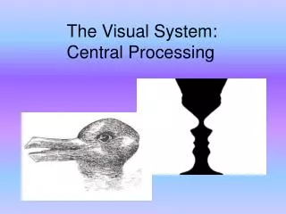
Organization of Cells in V1 • What sort of information are these cells detecting? • Is the information from any single cell in V1 informative? Anthony J Greene
Response Properties of Cells in V1 The extent to which columns will respond to stimuli with no interactions from other columns Cellular Activity Orientation Column Position on Occular Dominance Slab Anthony J Greene
Lateral Inhibition The column with the strongest response to a given stimuli will suppress the respondse of neighboring columns + - Anthony J Greene
Response Properties of Cells in V1 The extent to which columns will respond to stimuli with lateral inhibition from other columns Cellular Activity Orientation Column Position on Occular Dominance Slab Anthony J Greene
Processing at V1 Is Edge Detection Anthony J Greene
Edge Detection • While lateral inhibition normally improves the accuracy of edge detection, in this case it creates the “Deli Wall Illusion” Anthony J Greene
Understanding Acuity: Spatial Frequency Analysis • Measuring visual acuity: • Eye doctors use distance (e.g., 20/20) • Vision scientists use visual angle Anthony J Greene
Understanding Acuity: Spatial Frequency Anthony J Greene
Understanding Acuity: Spatial Frequency Anthony J Greene
Describing Processes in V1: Spatial Frequency Analysis Anthony J Greene
Describing Processes in V1: Spatial Frequency Analysis (cont.) • Orientation • Frequency • Contrast Decreasing Contrast Anthony J Greene
Spatial Frequency Analysis (cont.) • Fourier - French mathematician, came up with theory that one can create any complex wave through a summation of Sinusoids (or sub-parts, sub-waves) • Fourier Analysis divides all orientation specific cells in V1 according to the width of their receptive fields or Spatial Frequency • 1) Low • 2) Medium • 3) High Anthony J Greene
Spatial Frequency Analysis (cont.) • Neurons can then be viewed as Spatial Filters which separately analyze differing levels of detail or scale • Any scene can then be decomposed into images with varying spatial frequencies - low frequency images are blurry and only the most prominent features are represented - high frequency images exaggerate the fine details • Construing form vision in terms of an emergent property of these different scales of receptors is referred to as the Multichannel Model Anthony J Greene
Spatial frequency Analysis (cont.) Anthony J Greene
Spatial frequency Analysis (cont.) Anthony J Greene
Spatial frequency Analysis (cont.) • Once divided by width, cells can further be grouped according to their orientation specificity • This allows a vastly simplified organization of neural activity - 3 major variables - Spatial Frequency, Orientation & Contrast • Additionally, Fourier analysis helps explain how individual cells may contribute information to the aggregate Anthony J Greene
Spatial frequency Analysis (cont.) Anthony J Greene
Spatial frequency Analysis (cont.) Anthony J Greene
Spatial Frequency Analysis (cont.) • 1f gives the fundamental waveform • 2f ... xf : are called harmonics - increasing details Anthony J Greene
Spatial Frequency Analysis (cont.) Anthony J Greene
Spatial Frequency Illusions Anthony J Greene
Spatial Frequency Illusions Anthony J Greene
Spatial Frequency Illusions Anthony J Greene
Spatial Frequency Illusions Anthony J Greene
ColoratV1 • Among cells selective for orientation are patches of cells selective for color (and not orientation), which are known as Blobs. • Other cell (orientation specific cells) regions are known as interblobs. Anthony J Greene
Organization of V2 • Thin Stripes receive information from Blobs and pass it to V4 • Thick Stripes recieve information from complex cells and send it to V5 and V3 • Interstripes recieve information from simple cells and send it to V3 and V4 • Information at V2 is 3-D Anthony J Greene
Nerve or Cortical Damage 1) Retina / Optic Nerve 2) Optic Chiasm 3) Optic Tract 4) V1/V2 Anthony J Greene
Nerve or Cortical Damage Retina/Optic Nerve: Monocular blindness Anthony J Greene
Nerve or Cortical Damage Optic Chiasm: Nasal field blindness Anthony J Greene
Nerve or Cortical Damage Optic Chiasm: Bitemporal field blindness Anthony J Greene
Nerve or Cortical Damage Optic Tract/LGN/Radiations: Homonymous Blindness Anthony J Greene
Nerve or Cortical Damage V1: Quadrantic blindness Anthony J Greene
Nerve or Cortical Damage V1/V2: • Scotoma • Complete blindness • case of Blindsight Anthony J Greene
Higher Visual Areas • V3: Form & Dynamic Form • V4: Color • V5: Motion • IT: What System: Object Recognition • Lingual Gyrus of IT: Face Recognition • PP: Where System: Object Location and Navigation Anthony J Greene
Simplified Functional Visual Anatomy Anthony J Greene