Download
1 / 14
210 likes | 781 Views
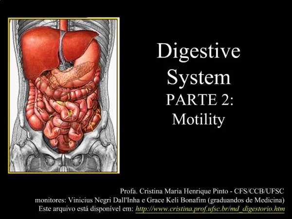
Lab # 7. Digestive System-2 . Liver and Gallbladder. Gross Anatomy of Liver. The liver is a reddish brown gland located immediately inferior to the diaphragm. 1-. It produces the bile to emulsify the fats. It is the body’s largest gland weighing about 1.4 kg (3 pounds). 2-.

E N D
Lab # 7 Digestive System-2
Liver and Gallbladder Gross Anatomy of Liver The liver is a reddish brown gland located immediately inferior to the diaphragm. 1- It produces the bile to emulsify the fats. It is the body’s largest gland weighing about 1.4 kg (3 pounds). 2- It processes nutrients from the blood. 3- It has many other metabolic and synthetic roles. Liver Functions: (a) Anterior view Round ligament Caudate lobe Inferior vena cava Right lobe Left lobe Falciform ligament It separates the right and left lobes and attaches the liver to the diaphragm and the abdominal wall. Gallbladder It marks the path of the fetal umbilical vein. Quadrate lobe (b) Posterior view
It separates the right and left lobes and attaches them to the abdominal wall. Falciform ligament Gastrosplenic ligament It attaches the stomach to the spleen. Left lobe Liver Lesser omentum Stomach Right lobe It attaches the stomach to the liver. Spleen
The Liver Functions: 1- It produces the bile to emulsify the fats 2- It processes nutrients from the blood 3- It has many other metabolic and synthetic roles Inferior vena cava Caudate lobe Caudate lobe Inferior vena cava Right lobe Left lobe Left lobe Right lobe Falciform ligament Anterior-superior view It stores and concen-trates the bile. Quadrate lobe Gallbladder Posterior-inferior view
Macroscopic Anatomy of Liver To the hepatic vein To the inferior vena cava Hepatic Lobule Central vein Hepatic Triad: Branch of hepatic portal vein Branch of proper hepatic artery Bile ductule To the right and left hepatic ducts Blood from the intestine and stomach Hepatocytes Bile canaliculum Hepatic sinusoid
The Hepatic Portal System The hepatic portal system drains all the blood from the abdominal digestive tract, as well as from the pancreas, gallbladder and spleen. Gastric vv. Hepatic portal v. Hepatic veins Splenic v. Inferior vena cava The hepatic portal system gives the liver first claim to the nutrients before the blood is distributed to the rest of the body. Superior mesenteric v. Inferior mesenteric v. It also allows the blood to be cleansed of bacteria and toxins picked up from the intestine.
Hepatic sinusoids: • They are blood-filled channels that fill spaces between the plates of hepatocytes. • Hepatic sinusoids are lined by a fenestrated endothelium that separates hepatocytes from blood cells and allows plasma into the space between the hepatocytes and endothelium. • Hepatocytes have brush border of microvilli that project into this space. • Hepatic macrophages (Kupffer cells) are phagocytic cells in the sinusoids that remove bacteria and debris from the blood. Functions of the hepatocytes: After a meal, the hepatocytes absorb from the blood: glucose, amino acids, iron, vitamins, and other nutrients for metabolism or storage. They remove and degrades hormones, toxins, bile pigments, and drugs. They secrete into the blood: albumin, lipoproteins, clotting factors, angiotensinogen, and other products. Between meals, they breaks down stored glycogen and releases glucose into the blood.
Liver Histology Bile duct Portal triad Central vein Hepatic artery proper Hepatic portal vein Hepatic or liver lobule Central vein Sinusoids Portal triad Sinusoids Bile canaliculi Hepatocytes
Liver Histology Sinusoids Hepatocytes Bile canaliculi Central vein Bile duct Portal triad Hepatic portal vein Hepatic artery proper
Hepatocyte Bile canaliculi Kupffer cells
Bile canaliculum Right hepatic ducts Bile ductule Left hepatic ducts Common hepatic duct Cystic duct Common bile duct Pancreatic duct Gallbladder Accessory pancreatic duct It stores and concentrates bile Pancreas Tail Body Duodenum Minor duodenal papilla Hepatopancreatic sphincter Head Major duodenal papilla Jejunum Hepatopancreatic ampulla Pancreas: It produces the pancreatic juice, which contains buffers and digestive enzymes (pancreatic alpha-amylase, pancreatic lipase, nucleases, and proteolytic enzymes)
Endocrine pancreas Exocrine pancreas Histology of the Pancreas Islets of Langerhans Pancreatic acini 1- Beta cells: Insulin Theyproduce the pancreatic juice, which contains buffers and digestive enzymes (pancreatic alpha-amylase, pancreatic lipase, nucleases, and proteolytic enzymes) Glucagon 2- Alpha cells: 3- Delta cells: Somatostatin 4- F cells: Pancreatic polypeptide
Bile and Pancreatic Ducts Model A Model B Cystic duct Cystic duct Common hepatic duct Gallbladder Common hepatic duct Common bile duct Common bile duct Accessory pancreatic duct Duodenal papilla Gallbladder Accessory pancreatic duct Common bile duct Duodenal papilla Pancreatic duct (duct of Wirsung) Duodenal ampulla Pancreatic duct (duct of Wirsung)
Histology of the Pancreas Pancreatic acini Pancreatic acini (seen in section) Islet of Langerhans (seen in relief) Capillary bed Connective tissue framework Pancreatic acini Pancreatic acini (seen in section) Islet of Langerhans (seen in section) Intralobular duct