Download
1 / 21
210 likes | 406 Views
Constructing Hexahedral Meshes of Abdominal Aortic Aneurysms for Use in Finite Element Analysis. Rowena Ong Vanderbilt University Mentor: Kara Kruse Computational Sciences and Engineering Division August 11, 2004. Introduction: What are abdominal aortic aneurysms (AAAs)?.

E N D
Constructing Hexahedral Meshes of Abdominal Aortic Aneurysms for Use in Finite Element Analysis Rowena Ong Vanderbilt University Mentor: Kara Kruse Computational Sciences and Engineering Division August 11, 2004
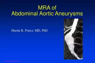
Introduction: What are abdominal aortic aneurysms (AAAs)? • Ballooning of aorta caused by weakened vessel walls • If untreated, vessel walls may rupture • AAAs 13th leading cause of death in U.S. • Mortality rate ~80% for ruptured AAAs
Introduction: What does AAA modeling involve? • Using equations to model wall stress and/or fluid flow through AAA • Three basic parts: • Equations modeling fluid flow and/or wall stress • 3D reconstruction of AAA from CT scans • Finite element analysis to numerically solve equations Cassada, Barnes, & Lilly, 2003
Introduction: Challenges of AAA Modeling Iliac bifurcation • Current finite element models of AAAs do not include both iliac bifurcation and thrombus • Iliac bifurcation and thrombus may play significant role in AAA formation
Introduction: Challenges of AAA Modeling • Generation of high-quality finite element mesh difficult at bifurcation • Why? • Finite element modeling – divide object into finite elements (blocks or tetrahedra) • Irregular geometry at bifurcation, difficult to generate regularly shaped elements • Important because irregularly shaped elements can yield inaccurate results (Di Martino et al., 2001)
Introduction • Goal of this project: Develop a semi-automatic method for generating high-quality meshes of AAAs • Including iliac bifurcation and thrombus • To be used in finite element analysis Enable researchers to explore effects of the iliac bifurcation and thrombus on AAA formation
Methods Thrombus Lumen Segment 1. Segment CT scans and generate triangular surface meshes using Amira Outer surface Inner surface Generate surfaces Obtain CT scans of AAA
Methods 2. Antiga and Steinman’s method (2004) and program for blood vessel surface generation was extended for AAAs a. Find centerlines • Compute Voronoi diagram, approximation of medial axis • Compute solution to Eikonal equation ∇ T = 1 / R(x) over Voronoi diagram using fast marching algorithm • Backtrace along path of steepest descent to find centerlines Antigua, Ene-Iordache, 2003 Antigua & Steinman, 2004
Methods b. Decompose bifurcation into 3 parts c. Build parametric mapping of the surface Longitudal mapping • Solve Δ f = 0 (where Δ is LaPlace-Beltrami operator) • Solve using finite element method Circumferential mapping • Use angular position of points with respect to the centerline Implemented in Visualization Toolkit (VTK) using Visual C++.NET Antigua & Steinman, 2004
Methods 3. Build hexahedral mesh of thrombus and lumen using sweeping algorithm
Results From actual patient data Outer and inner surfaces generated CT scans segmented
Results: Voronoi diagrams Inner surface Outer surface
Results: Centerlines Inner surface Outer surface
Results: Splitting lines Inner surface Outer surface
Results: Parametric representation Inner surface Outer surface
Results: Parametric Representation Close-up of bifurcation
Accomplishments • Extended Antiga and Steinman’s code to generate parameterized surfaces for AAAs • Program generates surfaces semi-automatically and objectively • Parameterized surfaces generated for real patient data
Future Work • Eliminate triangular area between patches in bifurcation region • Generate volumetric mesh from inner and outer surfaces • Write script to export meshes to Rhino or finite element modeling software • Build GUI for program
Long-term benefits • Program will allow high-quality finite element meshes to be generated semi-automatically and objectively • Will enable exploration of how iliac bifurcation and thrombus affect AAA formation through finite element modeling • Give physicians another tool to help evaluate AAA rupture risk
Acknowledgements Research Mentor Kara Kruse, M.S.E. Thanks to the following for AAA data: David C. Cassada, M.D., Michael B. Freeman, M.D., Mitchell H. Goldman, M.D. UT Medical Center, Dept. of Vascular Surgery Stephanie Barnes, Jennifer Lilly This work was funded by Mathematical, Information, & Computational Sciences DivisionOffice of Advanced Scientific Computing ResearchU.S. Department of Energy Through the Research Alliance for Math and Science (RAMS)
References • Antiga, L. and D. Steinman. 2004. Robust and Objective Decomposition and Mapping of Bifurcating Vessels. IEEE Trans. on Medical Imaging, 23(6):704-713. • Antiga, L., B. Ene-Iordache, A. Remuzzi. 2003. Centerline Computation and Geometric Analysis of Branching Tubular Surfaces with Application to Blood Vessel Modeling. 11th International Conference in Central Europe on Computer Graphics, Visualization and Computer Vision, 2003. • Antiga, L. 2003. Patient-Specific Modeling of Geometry and Blood Flow in Large Arteries. PhD Thesis. • Angenent, S., S. Haker, A. Tannenbaum, & R. Kikinis. 1999. On the Laplace–Beltrami Operator and Brain Surface Flattening. IEEE Trans. on Medical Imaging, 18(8):700-711.