Download
1 / 27
710 likes | 1.34k Views
Menopause. Danforth's Obstetrics and Gynecology, 10th Edition Dr Afsar tabatabai. Introduction. Human follicles begin their development during the fourth gestational month.

E N D
Menopause Danforth's Obstetrics and Gynecology, 10th Edition Dr Afsartabatabai
Introduction • Human follicles begin their development during the fourth gestational month. • Approximately 1,000 to 2,000 germ cells migrate to the gonadal ridge and multiply, reaching a total of 5 to 7 million around the fifth month of intrauterine life. • At this point, replication stops and follicle loss begins, declining to approximately 1 million by birth. • . In the female, between 12 and 18 weeks gestation, the germ cells enter meiosis and differentiate. • Over time, the population of oocytes will be depleted, without regeneration, through recruitment and apoptosis until fewer than a thousand oocytes exist and menopause ensues.
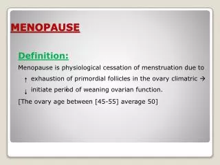
introduction • Menopause occurs at a median age of 51.4 years, with the age range in normal women being 42 to 58 years. • Approximately 90% of women experience menopause during the early 50s. • The other 10% of women experience menopause prior to 46 years of age (often termed early menopause), • with 1% of women experiencing menopause at an age younger than 40 years (premature menopause or premature ovarian failure )
Introduction • The age of menopause appears to be determined largely by genetics. • It does not appear to be related significantly to race, parity, height, weight, socioeconomic status, or age at menarche. • Evidence suggests that genetic and environmental factors influence the age of menopaus.
Reproductive Stages • Reproductive aging is a continuum beginning in utero and ending with menopause. • The staging system takes into account menstrual cyclicity, endocrinology, and symptomatology, beginning with menarche and ending with a woman's demise.(STRAW) • The foundation of the staging system is the final menstrual period (FMP). • Five stages precede the FMP and two follow it, for a total of seven stages. Stages -5 to -3 are called the reproductive interval, stages -2 to -1 are termed the menopausal transition, and stages +1 and +2 are known as the postmenopause
Reproductive Stages • The menopausal transition begins with variation in the menstrual cycle length (>7 days different from normal) in a woman with an elevated follicle-stimulating hormone (FSH) level. This stage ends with the FMP, which cannot be determined conclusively until after 12 months of amenorrhea. • Early postmenopause is defined as the first 5 years following the FMP. Late postmenopause is variable in length, beginning 5 years after the FMP and ending with the woman's death.
Endocrinology • The entire endocrine system in women changes with advancing age. • The somatotrophic axis begins to decline during the fourth decade, prior to the loss of ovarian function. • This decline in growth hormone is accelerated during ovarian failure and may, itself, accelerate the ovarian failure. • Although the thyroid gland undergoes progressive fibrosis with age and concentrations of T3 decline by 25% to 40%, elderly women remain clinically euthyroid.
Endocrinology • during the late fourth decade, FSH levels begin to rise even when cyclic menses continue(The most likely cause is a decrease in functional granulosa cells from the oocyte pool, with a decrease in inhibin B negative feedback ) • gradual declines in estradiol concentrations. • Dehydroepiandrosterone (DHEA) and its sulfated conjugate, DHEAS, have been shown to decrease with aging(Evidence suggests that physiologic levels of DHEA may protect against neoplasia, enhance insulin action, protect against osteoporosis, increase immune competency, and offer some cardioprotection. )
Systemic Effects of Declining Ovarian Function • The endometrium becomes atrophic • the uterus decreases in size • The postmenopausal vagina, devoid of estrogen treatment, becomes smaller in both length and caliber. • decreased elasticity of the vaginal wall. • The fallopian tubes contain both ciliated and secretory components. After age 60, cilia begin to disappear in the isthmic region, although they remain until a very old age in the ampulla and infundibulum. • With cessation of the production of estrogen and progesterone, glandular, ductal, and stromal involution occurs in mammary glamds. Connective tissue of the lobule becomes indistinguishable from other types of connective tissue. There is an accumulation of adipose tissue in the breast.
Menopausal Syndrome • many symptoms associated with aging in women are due to estrogen deficiency, but the decline in adrenal androgens and growth hormone may contribute. • definitely a result of estrogen deprivation include :vasomotor symptoms and urogenital atrophy • largely due to estrogen deficiency, but may be exacerbated by the relative growth hormone decline: Osteoporosis is likely, atherosclerotic CVD and psychosocial symptoms including insomnia, fatigue, short-term memory loss, and depression. • Both DHEAS and growth hormone may well have an impact on these age-related symptoms.
Vasomotor Symptoms A hot flash usually is characterized by intense warmth, that usually begins in the head, neck, and thorax and can spread in waves over the entire body. It may be preceded by pressure in the head and may be accompanied by heart palpitations. The length of the episode varies from seconds to approximately 5 minutes, although episodes as long as 30 minutes have been described. The event frequency varies from a few per year to 30 per day. Hot flashes result from estrogen deficiency. They occur in 65% to 76% of women who undergo spontaneous menopause or surgical oophorectomy.
Vasomotor Symptoms • For most women, the hot flashes commence prior to the FMP, although initially this may be perceived only as a sleep disturbance. • In general, the episodes are noted more frequently at night, and the dysfunctional sleep pattern that follows may result in fatigue, irritability, loss of concentration, and depression • More than 80% of women who experience hot flashes will experience them for longer than 1 year. Without treatment, the symptoms usually subside slowly over 3 to 5 years.
Vasomotor Symptoms • Treatment Estrogen therapy, in either oral or transdermal form, has a greater than 95% efficacy for the treatment of hot flashes For patients with contraindications to estrogen use, vasomotor symptoms can be treated with progestins, α2-adrenergic agonists (clonidine, methyldopa, lofexidine) and antidepressants (selective serotonin reuptake inhibitors [SSRIs], venlafaxine hydrochloride [Effexor]). The SSRI/selective norepinephrine reuptake inhibitors (SSNIs) and gabapentin appear to show modest reduction in hot flashes when compared with placebo
Genitourinary Atrophy • decrease in circulating estrogen levels has deleterious effects on urogenital epithelium. Up to 50% of postmenopausal women experience symptoms of vaginal atrophy. • The most common symptoms include dryness, irritation, itching, burning, and dyspareunia. • Atrophic vaginitis is associated with a rise in vaginal pH, which can lead to more frequent infections and worsening, irritative symptoms.
Genitourinary Atrophy • Estrogen replacement therapy (ERT) is an effective treatment for vaginal atrophy. • topical therapy by means of creams or vaginal rings may be advisable to limit systemic absorption. • local estrogen therapy can be used effectively to treat urogenital atrophy. • Vaginal estrogen cream or tablets can be used daily for approximately 2 to 3 weeks and then twice weekly after initial symptoms have improved and vaginal vascularization (hence, hormone uptake) has increased. • Vaginal estrogen frequently will improve symptoms of urinary frequency, dysuria, urgency, and postvoid dribbling.
Urinary Tract Infections • Recurrent UTIs in healthy postmenopausal women are associated with urinary incontinence, cystocele, and increased postvoid residual volumes. Other significant risk factors include at least one episode of UTI prior to menopause, urogenital surgery, and reduced urinary flow. • Changes in the vaginal environment after menopause also may predispose a woman to UTI. • These alterations include the absence of lactobacilli, elevated vaginal pH, and increased rate of vaginal colonization with Enterobacteriaceae. • The intravaginal administration of estrogen has been shown to reduce the rate of recurrent UTI by normalizing the vaginal environment.
Osteoporosis • Primary osteoporosis usually affects women between the ages of 55 and 70 years. • Approximately 30% of postmenopausal women have osteoporosis. • Secondary osteoporosis is caused by a specific disease (such as hyperparathyroidism) or medication usage (glucocorticoids, thyroid hormone excess, anticonvulsants). • The most common sites include the vertebrae and the long bones of the arms and legs. • Menopausal bone loss begins before the FMP during stage -1.
Osteoporosis • Bone loss following natural menopause is approximately 1% to 2% per year, compared with 3.9% per year following oophorectomy. • A woman's genetic background, lifestyle, dietary habits, and coexisting disease will impact the development of osteoporosis. • Cigarette smoking, caffeine use, and alcohol consumption are associated with increased bone loss, while weight-bearing activity appears to slow it. • The World Health Organization has defined osteoporosis as a hip BMD value, as measured by dual x-ray absorptiometry (DEXA), that is greater than 2.5 standard deviations below the adult peak (mean level for young, white women: t-score). • Women with existing fractures, regardless of BMD, are also classified as osteoporotic.
Osteoporosis • Treatment Hormone Replacement Therapy (prevention of osteoporosis and is FDA approved ) decreased fracture risk has been an important topic of research These studies have also reemphasized known risks of HRT, including pulmonary embolus, stroke, deep vein thrombosis, and gallbladder disease. HRT benefits include reduction in hot flash frequency and severity, improvement of atrophic vaginitis and UTIs, and prevention of osteoporosis and fractures. decreased risk of colorectal cancer.
Osteoporosis Calcitonin Intranasal calcitonin has been shown to improve spinal bone density and decrease the vertebral fracture rate in women with established osteoporosis. Bisphosphonates pamidronate, alendronate, and risedronate Raloxifene selective estrogen receptor modulators (SERMs) increases bone mass density in the spine and femoral neck and reduces the risk of vertebral fracture by 30% to 50% over no treatment. Calcium and Vitamin D 1,500 mg and 400 to 800 IU of vitamin D per day is probably sufficient to reduce the risk of fragility fractures by about 10%.
Osteoporosis Conclusions • An evidence-based approach is to recommend : appropriate calcium and vitamin D intake, smoking cessation, weight-bearing exercise, moderation in alcohol consumption, and fall prevention. If pharmacologic therapy is indicated, one of the above FDA-approved regimens should be instituted An alternative (complementary) approach to prevention is to focus on intervention beginning in childhood and adolescence, with attention to achieving maximal peak bone mass and minimizing premenopausal and postmenopausal bone loss.
Cardiovascular Disease • For women, CVD is largely a disease of postmenopause. • Women will now spend more than one third of their lives beyond menopause. • A woman's risk of heart disease is far lower than a man's risk until after menopause. • Randomized, controlled studies have failed to support a protective role for HRT. • There is no evidence from well-designed prospective trials supporting a role for either ERT or HRT for the primary indication of cardiovascular protection.
American Heart Association Recommendations • Secondary Prevention • HRT should not be initiated for the secondary prevention of CVD. • Primary Prevention • There are insufficient data to suggest that HRT should be initiated for the sole purpose of primary prevention of CVD.
Breast Cancer • The most compelling reason to believe that long-term use of postmenopausal estrogen increases the risk of breast cancer is the inherent biologic plausibility. • Many of the risk factors associated with breast cancer are thought to be linked to increased duration of exposure to estrogen over a woman's lifetime. • These risk factors include early menarche, late menopause, nulliparity, and older age at the birth of her first child. • The association of estrogen therapy and breast cancer remains controversial
This analysis reached the following conclusions: • Ever users of postmenopausal hormones had an overall increased relative risk of breast cancer of 1.14. • Current users for 5 or more years had a relative risk of 1.35 (CI 1.21 to 1.49), and the risk increased with increasing duration of use.