Download
1 / 32
720 likes | 1.55k Views
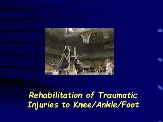
Knee Rehabilitation. Anatomy Review . Bony Anatomy Lower Leg Tibia Fibula Upper Leg Femur Patella. Anatomy Review. Lower Leg Musculature Anterior Tibialis Anterior Medial Tom, Dick and Harry Tibialis Posterior Extensor Digitorum Longus Extensor Hallicus Longus Lateral

E N D
Anatomy Review • Bony Anatomy • Lower Leg • Tibia • Fibula • Upper Leg • Femur • Patella
Anatomy Review • Lower Leg Musculature • Anterior • Tibialis Anterior • Medial • Tom, Dick and Harry • Tibialis Posterior • Extensor Digitorum Longus • Extensor Hallicus Longus • Lateral • Peroneals • Posterior • Gastrocnemius • Soleus • Tibialis Anterior
Anatomy Review • Thigh Musculature • Anterior • Quadriceps Femoris • Vastus Lateralis • Vastus Medialis • Vastus Intermedius • Rectus Femoris • Posterior • Biceps Femoris • Long Head • Short Head • Semi-tendonosis • Semi-membranosis • Gracilis
Anatomy Review • Ligaments • Medial Collateral • Lateral Collateral • Anterior Cruciate • Posterior Cruciate
Anatomy Review • Cartilage • Medial Meniscus • Lateral Meniscus • Articular Cartilage
Anatomy Review • Joint Capsule
Anatomy Review • Bursae
Knee Evaluation (History) • Determining the mechanism of injury is critical • History- Current Injury • Past history • Mechanism- what position was your body in? • Did the knee collapse? • Did you hear or feel anything? • Could you move your knee immediately after injury or was it locked? • Did swelling occur? • Where was the pain • History - Recurrent or Chronic Injury • What is your major complaint? • When did you first notice the condition? • Is there recurrent swelling? • Does the knee lock or catch? • Is there severe pain? • Grinding or grating? • Does it ever feel like giving way? • What does it feel like when ascending and descending stairs? • What past treatment have you undergone?
Knee Evaluation (Observation) • Observation • Walking, half squatting, going up and down stairs • Swelling, ecchymosis, • Leg alignment • Genu valgum and genu varum • Hyperextension and hyperflexion • Patella alta and baja • Patella rotated inward or outward • May cause a combination of problems
Knee Evaluation (Observation) • Knee Symmetry or Asymmetry • Do the knees look symmetrical? Is there obvious swelling? Atrophy? • Leg Length Discrepancy • Anatomical or functional • Anatomical differences can potentially cause problems in all weight bearing joints • Functional differences can be caused by pelvic rotations or mal-alignment of the spine
Palpation – Bony Medial tibial plateau Medial femoral condyle Adductor tubercle Gerdy’s tubercle Lateral tibial plateau Lateral femoral condyle Lateral epicondyle Head of fibula Tibial tuberosity Superior and inferior patella borders (base and apex) Around the periphery of the knee relaxed, in full flexion and extension Knee Evaluation (Palpation)
Palpation - Soft Tissue Vastus medialis Vastus lateralis Vastus intermedius Rectus femoris Quadriceps and patellar tendon Sartorius Medial patellar plica Anterior joint capsule Iliotibial Band Arcuate complex Medial and lateral collateral ligaments Pes anserine Medial/lateral joint capsule Semitendinosus Semimembranosus Gastrocnemius Popliteus Biceps Femoris Knee Evaluation (Palpation)
Knee Evaluation (Special Tests) • Active / Passive Range of Motion • Flexion – 0o to 135o • Extension – 130o to 0o • Manual Muscle Testing • Five Point grading system • 5 = Complete ROM against gravity, with full resistance • 4 = Complete ROM against gravity, with some resistance • 3 = Complete ROM against gravity, with no resistance • 2 = Complete ROM, with gravity omitted • 1 = Some muscle contractility with no joint motion • 0 = No muscle contractility • Knee Flexion / Extension • Hip Flexion / Extension / Internal Rotation / External Rotation • Dorsiflexion / Plantar Flexion
Knee Evaluation (Special Tests) • Joint Instability • Medial Collateral Ligament Instability
Knee Evaluation (Special Tests) • Joint Instability • Lateral Collateral Ligament Instability
Knee Evaluation (Special Tests) • Joint Instability • Anterior Cruciate Ligament (Lachman’s Test) • Will not force knee into painful flexion immediately after injury • Reduces hamstring involvement • At 30 degrees of flexion an attempt is made to translate the tibia anteriorly on the femur • A positive test indicates damage to the ACL
Knee Evaluation (Special Tests) • Joint Instability • Anterior Cruciate Ligament (Ant. Drawer) • Drawer test at 90 degrees of flexion • Tibia sliding forward from under the femur is considered a positive sign (ACL) • Should be performed w/ knee internally and externally to test integrity of joint capsule
Knee Evaluation (Special Test) • Other ACL Stability Tests • Pivot Shift Test • Used to determine anterolateral rotary instability • Position starts w/ knee extended and leg internally rotated • The thigh and knee are then flexed w/ a valgus stress applied to the knee • Reduction of the tibial plateau (producing a clunk) is a positive sign • Jerk Test • Reverses direction of the pivot shift • Moves from position of flexion to extension • W/out and ACL the tibia will sublux at 20 degrees of flexion
Joint Stability Tests • Posterior Cruciate Ligament Stability • Posterior Sag Test (Godfrey’s test) • Athlete is supine w/ both knees flexed to 90 degrees • Lateral observation is required to determine extent of posterior sag while comparing bilaterally
Knee Evaluation (Special Tests) • Other Posterior Cruciate Ligament Tests • Posterior Drawer Test • Knee is flexed at 90 degrees and a posterior force is applied to determine translation posteriorly • Positive sign indicates a PCL deficient knee
Knee Evaluation (Special Tests) • Meniscal Pathology • McMurray’s Meniscal Test • Used to determine displaceable meniscal tear • Leg is moved into flexion and extension while knee is internally and externally rotated in conjunction w/ valgus and varus stressing • A positive test is found w/ clicking and popping response Medial Meniscus Testing
Knee Evaluation (Special Tests) • McMurray Test Continued Lateral Meniscus Test
Knee Evaluation (Special Tests) • Meniscal Pathology • Apley’s Compression Test • Hard downward pressure is applied w/ rotation • Pain indicates a meniscal injury • Apley’s Distraction Test • Traction is applied w/ rotation • Pain will occur if there is damage to the capsule or ligaments • No pain will occur if it is meniscal
Knee Evaluation • Palpation of the Patella • Must palpate around and under patella to determine points of pain • Patella Grinding, Compression and Apprehension Tests • A series of glides and compressions are performed w/ the patella to determine integrity of patellar cartilage
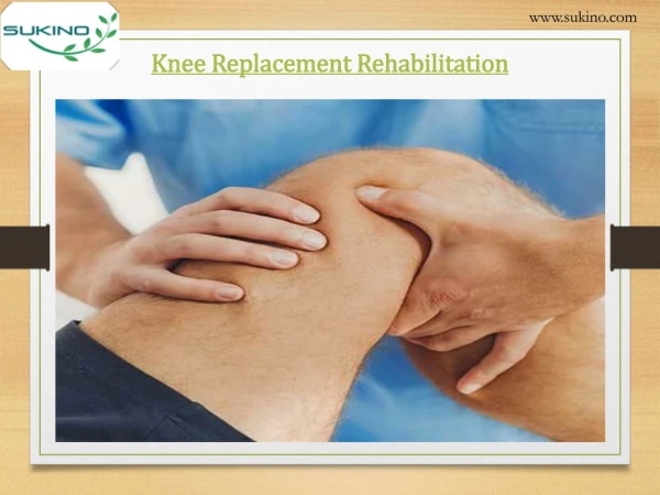
Knee Rehabilitation • Bag of Tricks • Range of Motion • Joint Mobilization, Soft-Tissue Mobilization • Neuromuscular Control • Proprioceptive Neuromuscular Facilitation • Postural Stability • Core Stability training • Muscular Strength, Endurance, and Power • Plyometrics, Open KC, Closed KC, Isokinetics, Aquatics • Cardiovascular Endurance
Knee Rehabilitation • Three simple keys • Range of Motion • Needed to increase motion and return to function as quickly as prudent and possible • Strength • Needed to deter further problems or protect the area of injury from further injury • Functionality • Needed to return the student-athlete or patient to normal daily activities within reason.
Knee Rehabilitation • Range of Motion Theory’s • Passive ROM is the key to early ROM • Active ROM starts and progresses as treatments continue • “Normal” Knee ROM • Knee Flexion = 0o to 130o+ • Knee Extension = 130o+ to 0o+
Knee Rehabilitation • Passive Range of Motion Exercises • Flexion Exercises • Wall Hangs (assisting device is gravity) • Towel Slides (assisting device is arms) • Stationary Bike (assisting device is other leg) • Extension Exercises • Table Hangs
Knee Rehabilitation • Strengthening • Closed Kinetic Chain • Used early in rehabilitation • More stable for the knee joint • Exercise include: • Mini-Squats (or with Swiss ball) • Wall Slides • Lunges (as ROM permits) • Leg Press Machine • Lateral Step-ups • T.K.E (Terminal Knee Extension) with T-Band
Knee Rehabilitation • Strengthening • Open Kinetic Chain • Also used early in rehabilitation • Exercise include: • Quad Sets • Hamstring Sets • Straight Leg Raises in four directions • Hamstring Curl Machine • Leg Extension Machine
Knee Rehabilitation • Functionality • Agility Drills / Training • Ladder • Dot Drills • Plyometric Drills / Training