Download
1 / 31
310 likes | 490 Views
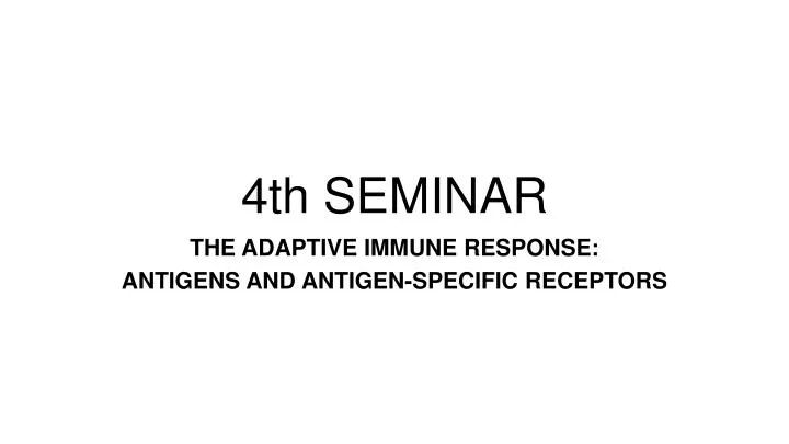
4th SEMINAR. THE ADAPTIVE IMMUNE RESPONSE: ANTIGENS AND ANTIGEN-SPECIFIC RECEPTORS. Cells Receptors. SENSING RECOGNITION. SENSING RECOGNITION. Signaling pathways Cell-Cell collaboration. SIGNALING. SIGNALING. Effector functions. RESPONSE. RESPONSE. DEFENSE SYSTEMS.

E N D
4th SEMINAR THE ADAPTIVE IMMUNE RESPONSE: ANTIGENS AND ANTIGEN-SPECIFIC RECEPTORS
Cells Receptors SENSING RECOGNITION SENSING RECOGNITION Signaling pathways Cell-Cell collaboration SIGNALING SIGNALING Effector functions RESPONSE RESPONSE DEFENSE SYSTEMS ADAPTIVE IMMUNITY INNATE IMMUNITY
RECOGNITION BY CELLS OF THE ADAPTIVE IMMUNE SYSTEM Antigen-specific receptors: B cell receptor (BCR) and T cell receptor (TCR) • The basic structure (90%) of the receptors (BCR or TCR) is common • Each cell expresses a receptor that is unique in specificity (the 10% difference means different specificity) • These differences in antigen-specificity are achieved during maturation in the central lymphoid organs (bone marrow and thymus)
ANTIGEN Any structure that can be recognized by the adaptive immune system (BCR, TCR). Antigenicity: ability of a chemical structure to bind specifically to a TCR or a BCR/antibody According to the results of the specific binding an antigen may be either: • Immunogenic: recognition induces an immune response • Tolerogenic: recognition induces tolerance (specific immune non-responsiveness)
FACTORS INFLUENCING IMMUNOGENICITY • Size(the bigger the better) • haptens: antigens that can not provoke an immune response because of their small size unless they are attached to a carrier molecule (e.g. a self peptide) • Genetics • Species (evolutionary the farther the better) • Individual (e.g. transplantation antigens) • Age(young: immature, old: decreasing number of lymphocytes) • Dose • Route(vaccination)subcutaneous > intravenous > oral / intranasalNot true for live vaccines (e.g. oral polio vaccine) • Adjuvant (vaccination) • substances that enhance the immune response to an antigen (aluminum salts, LPS, Freund’s adjuvant, TLR ligands) • depot effect – slower biodegradation, prolonged antigen intake by antigen presenting cells • activation of innate immunity • Physical status • corpuscle (cell, colloid) or soluble • denatured or native • Degradability antigen presentation by APCs
ANTIGENIC DETERMINANT (EPITOPE) Part of the antigen that directly interacts with the antigen binding site of the TCR or BCR/antibody.
B CELL EPITOPE T CELL EPITOPE Recognized by B cells proteins polysaccharides lipids DNA steroids etc. (many artificialmolecules) cell- or matrix-associated or soluble Recognized by T cells proteins mainly (8-23 amino acids) requires processing and presentation by APCs
L L L L H H H H b a signaling B CELL BCR AND ANTIBODY Antigen binding Membrane-bound Ig Antigen-specific Receptor (BCR) RECOGNIZING MOLECULE Secreted Ig Antigen-specific soluble protein EFFECTOR MOLECULE PLASMA CELL
IMMUNOGLOBULINS Definition: Glycoprotein molecules that are present on B cells as part of the BCR or produced by plasma cells as antibodies in response to an immunogenic antigen. Membrane-bound immunoglobulin (mIg) - BCR Secreted immunoglobulin (sIg) – antibody serum antibodies = gamma globulin fraction
STRUCTURE • 2x Heavy chain (light blue) • 2x light chain (dark blue) • Variable regions antigen binding • Constant regions disulfide bond carbohydrate CL VL CH2 CH3 CH1 hinge region VH
CDR3 150 100 CDR2 CDR1 variability index 50 FR2 FR1 FR4 FR3 0 25 75 100 50 aminoacid sequence N – C terminal HYPERVARIABLE REGIONS Light chain CDR2 CDR3 CDR1 Epitope CDR3 CDR1 CDR2 Heavy chain CDR = Complementarity Determining Region – those amino acids of the variable regions that directly interact with the epitope FR = frame – those amino acids of the variable regions that do not interact directly with the epitope (stabilizer function)
DIFFERENT VARIABLE REGIONS DIFFERENT ANTIGEN-BINDING SITE DIFFERENT SPECIFICITY
idiotype allotype isotype (Classes/subclasses) Sequence variability of H/L-chain constant regions Allelic variants Sequence variability of H and L-chain variable regions (individual, clone- specific)
HUMAN IMMUNOGLOBULIN CLASSESencodedbydifferentstructuralgenesegments (isotypes) • IgG - gamma (γ) heavy chains • IgM - mu (μ) heavy chains • IgA - alpha (α) heavy chains • IgD - delta (δ) heavy chains • IgE - epsilon (ε) heavy chains lightchaintypes • kappa (κ) • lambda (λ)
papain IMMUNOGLOBULIN FRAGMENTS STRUCTURE/FUNCTION RELATIONSHIPS • Fab • antigen binding • valence = 1 • specificity determined by VH and VL • Fc • effectorfunctions VH VL Fc Fab
pepsin Fc peptides F(ab’)2 IMMUNOGLOBULIN FRAGMENTS STRUCTURE/FUNCTION RELATIONSHIPS • F(ab’)2 - Bivalent!
ANTIBODY FUNCTION • Role of the Fab part: • Binds the antigen • May form crosslinks between antigens (precipitation / agglutination – see later) • Neutralization: binding can block the enzyme or toxin or other virulence factors of pathogens and can avoid damage to host cells • Role of the Fc part: • Activate cells carrying Fc-receptors on their surfaces: • Phagocytic cells – opsonized phagocytosis • NK cells – antibody-dependent cellular cytotoxicity (ADCC) • Activates the complement systemvia the classical pathway
OPSONIZATION NEUTRALIZATION
(A) High-affinity FcRs on the surface of the cell bind monomeric Ig before it binds to antigen. (mast cell) (B) Low-affinity FcRs bind multiple Igs that have already bound to a multivalent antigen. (macrophage, NK cell)
S S S S S S S S S S S S S S S S S S S S S S S S S S S S s s s s s s s s s s s s s s C C C C C C C J J J J J J J C C C C C C C S S S S S S S S S S S S S S C C C C C C C C C C C C C C pIgR and IgA are internalised S S S S S S S S S S S S S S C C C C C C C C C C C C C C Polymeric Ig receptors are expressed on the basolateral surface of epithelial cells to capture IgA produced in the mucosa B SECRETORY IgA AND TRANSCYTOSIS MUCUS ‘Stalk’ of the pIgR is degraded to release IgA containing part of the pIgR (the secretory component) IgA and pIgR are transported to the apical surface in vesicles Epithelial cell B cells located in the submucosaproduce dimeric IgA
TCR • Alpha and beta chains instead of light and heavy (innate subgroup of T cells express gamma-delta chains as TCR) • Both chains are membrane-bound • Monovalent interaction with the antigen • Antigen recognition requires presentation by antigen presenting cells via MHC molecules • Recognize peptide antigens (mainly) • No secreted form
ANTIGEN PRESENTATION • T cells that express CD8 as co-receptor (cytotoxic T cells) recognize peptides presented via MHC class I molecules • T cells that express CD4 as co-receptor (helper T cells) recognize peptides presented via MHC class II molecules • For activation both cell types require the help of APCs • Antigen presentations that lead to T cell activation take place in the secondary lymphoid tissues (e.g. lymph nodes) • MHC I is expressed by every nucleated cell RBCs don’t express them • Professional antigen presenting cells express MHC class I and class II molecules: • macrophage (innate) • DC (innate) • B cell (adaptive)
MHC RESTRICTION OF T CELL RECOGNITION • A given TCR recognizes a defined MHC – peptide complex • The same peptide presented by another MHC is not recognized by the same TCR • Another peptide bound to the same MHC is not recognized by the same TCR 1. 2. 3.
ε δ ε γ α β ζ ζ ACTIVATION OF TCR AND BCR ITAM Immunoreceptor Tyrosine-based Activation Motif ACTIVATION
SUPERANTIGENS Microbial proteins that bind to and activate all the T cells that express a particular set or family of TCR molecules. The activation is independent from the presented antigen. Leads to polyclonal T cell activation that causes life threatening inflammatory responses.
superantigen conventionalantigen polyclonal T cell response 1:4 - 1:10 SUPERANTIGENS monoclonal/oligoclonal T cell response 1:104 - 1:105 activated T cells 107 – 108 / 1011 1010 / 1011