Download
1 / 29
430 likes | 1.79k Views
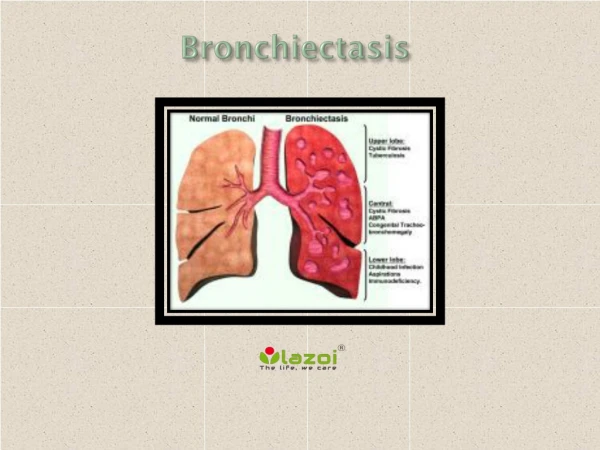
Bronchiectasis. Bronchiectasis is the term used to describe abnormal dilatation of the bronchi . It is usually acquired but may result from an underlying genetic or congenital defect of airway defences. Morphological types. Cylindrical or tubular bronchiectasis Varicose

E N D
Bronchiectasis is the term used to describe abnormal dilatation of the bronchi. It is usually acquired but may result from an underlying genetic or congenital defect of airway defences
Morphological types • Cylindrical or tubular bronchiectasis • Varicose • saccular or cystic bronchiectasis
Aetiology and pathogenesis of Bronchiectasis • Bronchiectasis is usually caused by chronic inflammation and infection in airways. • TB is the most common cause of Bronchiectasis world-wide. • Localised bronchiectasis may be due to bronchial distension resulting from the accumulation of pus beyond an obstructing bronchial lesion, such as enlarged tuberculous hilar lymph nodes, a bronchial tumour or an inhaled foreign body (e.g. an aspirated peanut).
CAUSES OF BRONCHIECTASIS • Congenital - Cystic fibrosis . - Ciliary dysfunction syndromes : Primary ciliary dyskinesia (immotile cilia syndrome) Kartagener's syndrome (sinusitis and transposition of the viscera) - Primary hypogammaglobulinaemia • Acquired-children - Pneumonia (complicating whooping cough or measles) - Primary TB . - Inhaled foreign body . Acquired-adults -Suppurative pneumonia. -Pulmonary TB . -Bronchial tumours - Allergic bronchopulmonary aspergillosis complicating asthma
Pathology • The bronchiectatic cavities may be lined by granulation tissue, squamous epithelium or normal ciliated epithelium. • There may also be inflammatory changes in the deeper layers of the bronchial wall and hypertrophy of the bronchial arteries. • Chronic inflammatory and fibrotic changes are usually found in the surrounding lung tissue.
Clinical features of bronchiectasis FEATURES OF BRONCHIECTASIS Due to accumulation of pus in dilated bronchi • Chronic productive cough usually worse in mornings and often brought on by changes of posture. • Sputum often copious and persistently purulent in advanced disease. • Halitosis is a common accompanying feature • SOB
Due to inflammatory changes in lung and pleura surrounding dilated bronchi • Fever, malaise and increased cough and sputum volume when spread of infection causes pneumonia, which may be associated with pleurisy. • Recurrent pleurisy in the same site often occurs in bronchiectasis
Haemoptysis Can be slight or massive and is often recurrent. Usually associated with purulent sputum or an increase in sputum purulence. Can, however, be the only symptom in so-called 'dry bronchiectasis' • General health When disease is extensive and sputum persistently purulent a decline in general health occurs with weight loss, anorexia, lassitude, low-grade fever, and failure to thrive in children. In these patients digital clubbing is common
Clinical features of bronchiectasis con… • Bronchiactasis may involve any part of the lungs but the more efficient drainage by gravity of the upper lobes usually produces less serious symptomes & complications than when bronchiactasis involves the lower lobes.
Physical signs in the chest may be unilateral or bilateral. • If the bronchiectatic airways do not contain secretions and there is no associated lobar collapse, there are no abnormal physical signs. • When there are large amounts of sputum in the bronchiectatic spaces numerous coarse crackles may be heard over the affected areas. • When collapse is present the character of the physical signs depends on whether or not the proximal bronchus supplying the collapsed lobe is patent (breath sounds are diminished if the airway is obstructed). • Advanced disease may lead to scarring with associated overlying bronchial breathing.
When to suspect bronchiectasis? Chronic cough, sputum hemoptysis Coarse rales Persistent respiratory symptoms Recurrent pneumonia Progressive obstructive lung disease clubbing
Sputum production Mild <15 cc/d Moderate 15-150 cc/d Severe >150 cc/d Clinical Characteristics
If wide spread Dyspnea Clubbing of the fingers h pulmonary blood pressure Cor pulmonale Bronchiectasis
Investigations • Bacteriological and mycological examination of sputum • In addition to common respiratory pathogens, sputum culture may reveal Pseudomonas aeruginosa, fungi such as Aspergillus and various Mycobacteria. • Frequent cultures are necessary to ensure appropriate treatment of resistant organisms. • Radiological examination Bronchiectasis, unless very gross, is not usually apparent on a chest X-ray. • In advanced disease, thickened airway walls, cystic bronchiectatic spaces, and associated areas of pneumonic consolidation or collapse may be visible. • CT scane of chest is much more sensitive, and shows thickened dilated airways
Assessment of ciliary function • A screening test can be performed in patients suspected of having a ciliary dysfunction syndrome by assessing the time taken for a small pellet of saccharin placed in the anterior chamber of the nose to reach the pharynx, when the patient can taste it. This time should not exceed 20 minutes and is greatly prolonged in patients with ciliary dysfunction. • Ciliary beat frequency may also be assessed using biopsies taken from the nose. • Structural abnormalities of cilia can be detected by electron microscopy.
Management • Postural drainage • Physiotherapy : Patients should be instructed how to perform regular daily physiotherapy to keep the dilated bronchi empty of secretions. Efficiently performed, this is of great value both in reducing the amount of cough and sputum and in preventing recurrent episodes of bronchopulmonary infection.
Patients should adopt a position in which the lobe to be drained is uppermost. Deep breathing followed by forced expiratory manoeuvres (the 'active cycle of breathing' technique) is of help in allowing secretions in the dilated bronchi to gravitate towards the trachea, from which they can be cleared by vigorous coughing. 'Percussion' of the chest wall with cupped hands may help to dislodge sputum, and a number of mechanical devices are available which cause the chest wall to oscillate, thus achieving the same effect. • The optimum duration and frequency of physiotherapy depends on the amount of sputum but 5-10 minutes once or twice daily is a minimum for most patients.
In patients with airflow obstruction, inhaled bronchodilators and corticosteroids should be used to enhance airway patency
Antibiotic therapy Antibiotics which can be used in patients with bronchiectasis are : amoxycillin , clarithromycin , Co-amoxyclav ….). Some present difficult therapeutic problems because of secondary infection with bacteria such as staphylococci and Gram-negative bacilli, in particular Pseudomonas species. In these circumstances antibiotic therapy should be guided by the microbiological results but frequently requires the use of oral ciprofloxacin (250-750 mg 12-hourly) or ceftazidime by intravenous injection or infusion (1-2 g 8-hourly).
Surgical treatment • Surgery is only indicated in a small proportion of cases. • These are usually young patients in whom the bronchiectasis is unilateral and confined to a single lobe or segment as demonstrated by CT. • Unfortunately, many of the patients in whom medical treatment proves unsuccessful are also unsuitable for pulmonary resection because of either extensive bronchiectasis or coexisting chronic lung disease. Resection of areas of bronchiectasis lung has no role in the management of the progressive form of bronchiectasis - for example those associated with ciliary dysfunction & cystic fibrosis .
Surgical treatment • Localised bronchiectasis • Proximal obstructive lesion • Massive hemoptysis • Recurrent infections
Prognosis • The disease is progressive when associated with ciliary dysfunction and cystic fibrosis, and eventually causes respiratory failure. • In other patients the prognosis can be relatively good if postural drainage is performed regularly and antibiotics are used judiciously.
Prevention • As bronchiectasis commonly starts in childhood following measles, whooping cough or a primary tuberculous infection, it is essential that these conditions receive adequate prophylaxis and treatment. • The early recognition and treatment of bronchial obstruction are also particularly important.
complications • 1-pneumonia , lung abscess. • 2-pleurisy , empyema • 3- sinusitis. • 4- massive haemoptysis. • 5- cor- pulmonale • 6- brain abscess. • 7- Amyloidosis. • 8- Emphysema.