Download
1 / 17
180 likes | 287 Views
Mock siRNA siRNA Day 3 Day 8 Day 3 Day 8 1 2 3 4. DNA-PKcs Beta actin. (A). (B). (C). *. *. *. *. Fig. 1. Fig 1.

E N D
Mock siRNA siRNA Day 3 Day 8 Day 3 Day 8 1 2 3 4 DNA-PKcs Beta actin (A) (B) (C) * * * * Fig. 1
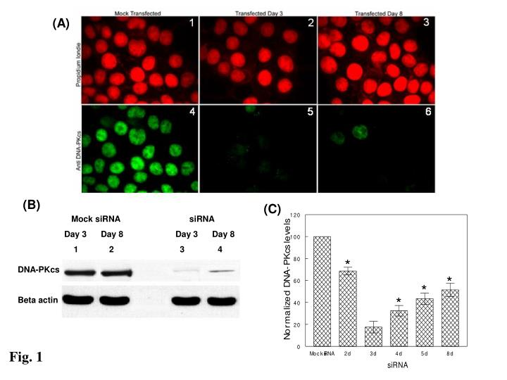
Fig 1 • Fig. 1 Silencing of DNA-PKcs expression in MCF-7 cells after transfection with siRNA targeted to knockdown the expressioin of Prkdc gene. (A) Immunofluorescence assay showing silencing of DNA-PKcs protein in siRNA DNA-PKcs double transfected MCF-7 cells. Bottom row (4-6), fluorescence microscope images were obtained on different days after the 2nd transfection (here showing day 3 and 8). Top row (1-3), red nuclear stain by propidium iodide. Cells on the bottom row were immunostained with FITC labeled mouse anti-DNA-PKcs (green); photographs were taken with FITC and propidium iodide filters. These data are representative of two independent experiments. (B) Western blotting showing reduced expression of DNA-PKcs protein in siRNA DNA-PKcs double transfected MCF-7 cells. MCF-7 cell lysates were prepared on different days after the 2nd transfection (here shown day 3 and day 8) from transfected and mock transfected (control siRNA only) cells. 50μg of protein was loaded in each lane and probed using rabbit polyclonal antibody against DNA-PKcs (~465 kDa) and mouse monoclonal antibody against beta actin (~44 kDa) used as standard. Lanes 1 and 2: mock transfected cells; lanes 3 and 4: transfected cells. (C) Quantitation of immunoblotting results. DNA-PKcs band total intensity of mock-transfected MCF-7 has been used for control and all values were normalized to that. Values are averages from two independent experiments. Error bars, SEM. * Statistical significant from 3d sample at p<0.05
(B) (A) Repair time (hrs) γ-H2AX NR 1 24 48 72 IC86621 Control MO59-J MO59-K (C) 100 100 90 MCF-7 MO59-J/K 90 80 80 70 70 60 60 DSBs/Gbp DSBs/Gbp 50 50 MCF7-IC86621 40 40 30 30 MCF7-siRNA 20 20 MO59-J MCF7-Control 10 10 MO59-K 0 0 0 8 16 24 32 0 8 16 24 32 40 48 56 64 72 80 Repair time (hrs) Repair time (hrs) Fig. 2
Fig. 2 • Fig. 2 Processing of DSBs in DNA-PKcs proficient and deficient cell lines (IC86621-, NU7026-, siRNA-MCF-7 and MCF-7, MO59-J and MO59-K) as a function of post-irradiation time. (A) Representative microscopic fluorescence images of γ-H2AX foci accumulation after 5 Gy IR at times (1, 24, 48 and 72 hrs). Non-irradiated samples (NR) are also shown. Nuclei were stained using either propidium iodide PI (MCF-7-top) or DAP1 (MO59-J/K-btm) (B) Quantitation of the normalized total mean γ-H2AX fluorescence intensity expressed as intensity/nucleus (% of maximum value at 1 hr). (C) Detection of DSB repair using PFGE analysis. Values for DNA-PK siRNA cells (up to 24 hrs) are taken from Peddi et al. [24] to allow direct comparison while for 48 and 72 hrs are from this study (crosses). Values are averages from three (MCF-7) or two (MO59-J/K) independent experiments. Inset: Average values for MCF-7 with repair times up to 12 hr. Small symbols, individual data points from independent irradiation experiments; Large symbols, averages. Error bars, SEM; in some cases are smaller than the corresponding symbol. Closed symbols, control MCF-7 or MO59-K cells. Closed large circles, NU7026-treated MCF-7. Open symbols, IC86621-treated MCF-7 cells or M059-J. Statistically significant differences between IC86621-/NU7026-MCF-7 and controls (*) and IC86621-/NU7026-MCF-7 and siRNA-MCF-7 (**) at p<0.05 are shown.
Figure 3. Persistence of phospho-Thr2609 DNA-PKcs foci until 5 days in 100 μM IC86621 pretreated MCF-7 cells after 5 Gy γ irradiation MCF-7 cells were incubated with 100 μM IC86621 (treated cells) or DMSO (control cells) for 24 h. The cells were irradiated with 5 Gy γ-rays. Following irradiation, the cells are returned to 37oC incubator. Cells were fixed at 0.5, 3, 12, 24, 48, 72 and 120 h after irradiation. Both control sample (DMSO treated only) and test samples (IC86621 treated) are immunostained with rabbit polyclonal to phospho-(Thr2609) DNA-PKcs antibody (green foci). DAPI is used to stain nucleus (Blue) in all cells. Photographs of the same cells were taken with FITC and DAPI filters and later were merged using Adobe Photoshop 7.0 software. Compared to the DMSO treated irradiated cells, the rate of disappearance of phospho-(Thr2609) DNA-PKcs is delayed in IC86621 treated cells. The results shown here are representative of two independent experiments.
Effect of DNA-PK deficiency on gamma-H2AX Foci PersistenceMO59-J: DNA-PK deficientMO59-K: DNA-PK proficient Alex Georgakilas NTUA
MO59-J Cell Controls (0 Gy) 0 hr 48 hr 24 hr 72 hr
MO59-K Cell Controls (0 Gy) 0 hr 48 hr 24 hr 72 hr
J Cells 3 Gy 0.5 hr 1 hr 3 hr 6 hr 10 hr 24 hr 48 hr 72 hr
K Cells 3 Gy 0.5 hr 1 hr 3 hr 6 hr 10 hr 24 hr 48 hr 72 hr
J Cells 5 Gy 0.5 hr 1 hr 3 hr 6 hr 10 hr 24 hr 48 hr 72 hr
K Cells 5 Gy 0.5 hr 1 hr 3 hr 6 hr 10 hr 24 hr 48 hr 72 hr
J vs. K cells 10 hr post 3 Gy IR J Cells K Cells
J vs. K cells 10 hr post 5 Gy IR J Cells K Cells
Conclusions • J cells (deficient in DNA-PK expression) have greater foci persistence over time after IR. • Overall numbers of foci between J cells and K cells at 30 min post-IR seem similar. • DNA-PK seems to play a role in foci removal.
Original western blots for the detection of XRCC1 for MCF-7 cells treated with ICC8621 inhibitor