Download
1 / 1
10 likes | 168 Views
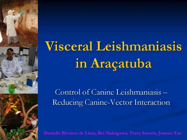
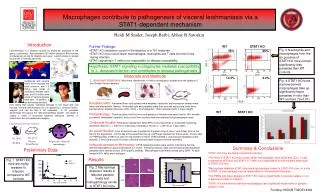
Harvested Spleen. Sick, infected hamster. Parasites Isolated. PBS DRUG. 10X. macrophage. Th2. IL-10. Th1. IFN g. Disease Exacerbation. Healing. Target cells = host cell macrophages. 40X.

E N D
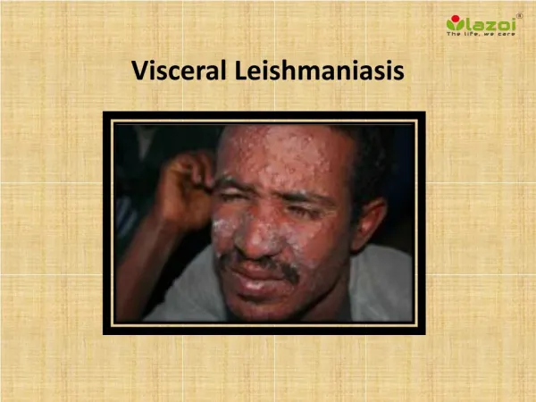
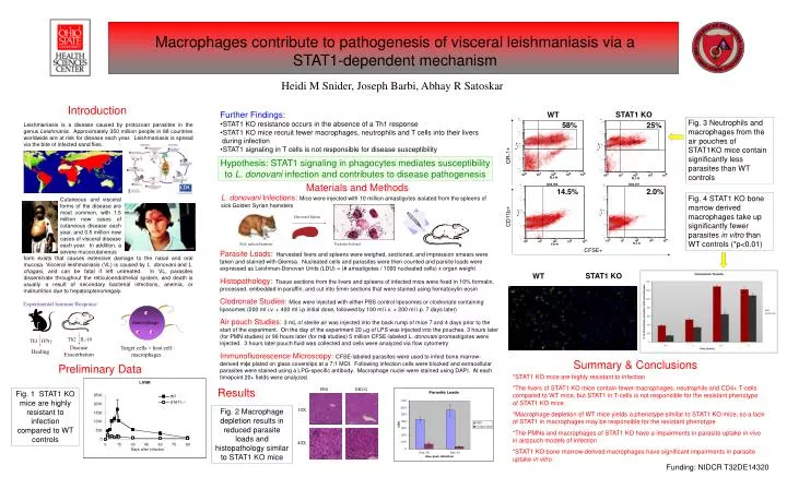
Harvested Spleen Sick, infected hamster Parasites Isolated PBS DRUG 10X macrophage Th2 IL-10 Th1 IFNg Disease Exacerbation Healing Target cells = host cell macrophages 40X Macrophages contribute to pathogenesis of visceral leishmaniasis via a STAT1-dependent mechanism * * * Heidi M Snider, Joseph Barbi, Abhay R Satoskar Introduction • Further Findings: • STAT1 KO resistance occurs in the absence of a Th1 response • STAT1 KO mice recruit fewer macrophages, neutrophils and T cells into their livers • during infection • STAT1 signaling in T cells is not responsible for disease susceptibility STAT1 KO WT Fig. 3 Neutrophils and macrophages from the air pouches of STAT1KO mice contain significantly less parasites than WT controls 58% 25% Leishmaniasis is a disease caused by protozoan parasites in the genus Leishmania. Approximately 350 million people in 88 countries worldwide are at risk for disease each year. Leishmaniasis is spread via the bite of infected sand flies. GR-1+ GR-1+ Hypothesis: STAT1 signaling in phagocytes mediates susceptibility to L. donovani infection and contributes to disease pathogenesis Materials and Methods 14.5% 2.0% L. donovani Infections: Mice were injected with 10 million amastigotes isolated from the spleens of sick Golden Syrian hamsters Fig. 4 STAT1 KO bone marrow derived macrophages take up significantly fewer parasites in vitro than WT controls (*p<0.01) Cutaneous and visceral forms of the disease are most common, with 1.5 million new cases of cutaneous disease each year, and 0.5 million new cases of visceral disease each year. In addition, a severe mucocutaneous CD11b+ CFSE+ Parasite Loads:Harvested livers and spleens were weighed, sectioned, and impression smears were taken and stained with Giemsa. Nucleated cells and parasites were then counted and parsite loads were expressed as Leishman-Donovan Units (LDU) = (# amastigotes / 1000 nucleated cells) x organ weight Histopathology:Tissue sections from the livers and spleens of infected mice were fixed in 10% formalin, processed, embedded in paraffin, and cut into 5mm sections that were stained using hematoxylin eosin Clodronate Studies: Mice were injected with either PBS control liposomes or clodronate containing liposomes (200 ml i.v. + 400 ml i.p initial dose, followed by 100 ml i.v. + 200 ml i.p. 7 days later) Air pouch Studies: 3 mL of sterile air was injected into the back rump of mice 7 and 4 days prior to the start of the experiment. On the day of the experiment 20 g of LPS was injected into the pouches. 3 hours later (for PMN studies) or 96 hours later (for mϕ studies) 5 million CFSE-labeled L. donovani promastigotes were injected. 3 hours later pouch fluid was collected and cells were analyzed via flow cytometry Immunofluorescence Microscopy: CFSE-labeled parasites were used to infect bone marrow-derived mϕs plated on glass coverslips at a 7:1 MOI. Following infection cells were blocked and extracellular parasites were stained using a LPG-specific antibody. Macrophage nuclei were stained using DAPI. At each timepoint 20+ fields were analyzed. form exists that causes extensive damage to the nasal and oral mucosa. Visceral leishmaniasis (VL) is caused by L. donovani and L. chagasi, and can be fatal if left untreated. In VL, parasites disseminate throughout the reticuloendothelial system, and death is usually a result of secondary bacterial infections, anemia, or malnutrition due to hepatosplenomegaly. STAT1 KO WT Experimental Immune Response Summary & Conclusions Preliminary Data • STAT1 KO mice are highly resistant to infection • The livers of STAT1 KO mice contain fewer macrophages, neutrophils and CD4+ T-cells compared to WT mice, but STAT1 in T-cells is not responsible for the resistant phenotype of STAT1 KO mice • Macrophage depletion of WT mice yields a phenotype similar to STAT1 KO mice, so a lack of STAT1 in macrophages may be responsible for the resistant phenotype • The PMNs and macrophages of STAT1 KO have a impairments in parasite uptake in vivo in airpouch models of infection • STAT1 KO bone marrow-derived macrophages have significant impairments in parasite uptake in vitro Results Fig. 1 STAT1 KO mice are highly resistant to infection compared to WT controls Fig. 2 Macrophage depletion results in reduced parasite loads and histopathology similar to STAT1 KO mice Funding: NIDCR T32DE14320