Download
1 / 1
10 likes | 125 Views
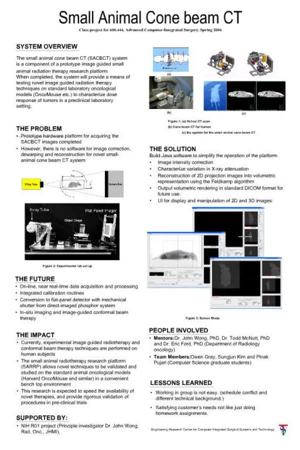
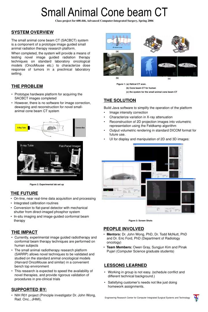
X-Ray Tube. Camera Box. Small Animal Cone beam CT Class project for 600.446, Advanced Computer-Integrated Surgery, Spring 2006. SYSTEM OVERVIEW The small animal cone beam CT (SACBCT) system is a component of a prototype image guided small animal radiation therapy research platform.

E N D
X-Ray Tube Camera Box Small Animal Cone beam CTClass project for 600.446, Advanced Computer-Integrated Surgery, Spring 2006 SYSTEM OVERVIEW The small animal cone beam CT (SACBCT) system is a component of a prototype image guided small animal radiation therapy research platform. When completed, the system will provide a means of testing novel image guided radiation therapy techniques on standard laboratory oncological models (OncoMouse etc.) to characterize dose response of tumors in a preclinical laboratory setting. (a) (b) (c) Figure 1: (a) Helical CT scan. (b) Cone beam CT for human (c) the system for the small animal cone beam CT THE PROBLEM • Prototype hardware platform for acquiring the SACBCT images completed • However, there is no software for image correction, dewarping and reconstruction for novel small-animal cone beam CT system THE SOLUTION Build Java software to simplify the operation of the platform • Image intensity correction • Characterize variation in X-ray attenuation • Reconstruction of 2D projection images into volumetric representation using the Feldkamp algorithm • Output volumetric rendering in standard DICOM format for future use. • UI for display and manipulation of 2D and 3D images: Figure 2: Experimental lab set up THE FUTURE • On-line, near real-time data acquisition and processing • Integrated calibration routines • Conversion to flat-panel detector with mechanical shutter from direct-imaged phosphor system • In-situ imaging and image-guided conformal beam therapy Figure 3: Screen Shots PEOPLE INVOLVED • Mentors: Dr. John Wong, PhD, Dr. Todd McNutt, PhD and Dr. Eric Ford, PhD (Department of Radiology oncology) • Team Members: Owen Gray, Sungjun Kim and Pinak Pujari (Computer Science graduate students) THE IMPACT • Currently, experimental image guided radiotherapy and conformal beam therapy techniques are performed on human subjects • The small animal radiotherapy research platform (SARRP) allows novel techniques to be validated and studied on the standard animal oncological models (Harvard OncoMouse and similar) in a convenient bench top environment • This research is expected to speed the availability of novel therapies, and provide rigorous validation of procedures in pre-clinical trials LESSONS LEARNED • Working in group is not easy. (schedule conflict and different technical background.) • Satisfying customer’s needs not like just doing homework assignments. SUPPORTED BY: • NIH R01 project (Principle investigator Dr. John Wong, Rad. Onc., JHMI), Engineering Research Center for Computer Integrated Surgical Systems and Technology