Download

1 / 71
740 likes | 1.24k Views
Preoperative Evaluation of the Patient with Pulmonary Disease. Gregory C. Kane, MD, FACP, FCCP Professor of Medicine, Division of Pulmonary Medicine and Critical Care Vice-Chairman for Education and Internal Medicine Residency Director Jefferson Medical College Philadelphia, PA.

E N D
Preoperative Evaluation of the Patient with Pulmonary Disease Gregory C. Kane, MD, FACP, FCCP Professor of Medicine, Division of Pulmonary Medicine and Critical Care Vice-Chairman for Education and Internal Medicine Residency Director Jefferson Medical College Philadelphia, PA
Preoperative Evaluation of the patient with Pulmonary Disease • Assessment tools • Who’s at risk? • Assessment of • Patients undergoing thoracotomy • Patients undergoing abdominal surgery • Outcomes after AAA repair (endovascular vs. open) • Non-thoracic procedures • Other procedures including obesity surgery • Special considerations • CABG in patients with COPD and reducing PPC • Measures to prevent post-op complications
Assessment tools • History • Functional status • Exam (presence/absence of Cor pulmonale) • Imaging • Spirometry • Special measures of lung function (ieDlCO) • ABG • Echocardiogram • Exercise testing
Patients at Risk • Chronic Lung Disease • Asthma • Smoking • Advanced Age • Obesity • General medical status • Upper respiratory infection
Patients at RiskACP Guidelines • Chronic Lung Disease (COPD) • Asthma • Smoking • Advanced Age > 60 • Obesity • General medical status (ASA Class II or >) • CHF • Functionally Dependant • Upper respiratory infection Annals Intern Med 2006; 144:575-580
Preoperative Evaluation of the patient with Lung Cancer • Anatomic Resectablity (surgical staging required) • Stage I • Stage II • Selected stage IIIa • Operability (physiologic resectibility) • Predicted Post-op FEV1> 0.8 liter • No CO2 retention • No pulmonary hypertension • VO2 max > 15ml/kg/min Batra, Kane, and Weibel Clin Pulm Med 2002; Miller J Thor Cardiovasc Surg 1993; Smtih etal Am Rev Resp Ds 1984
Staging- changes last decade (1997) • Stage I now divided into Ia (T1; < 3cm) and Ib (T2) • Stage II now IIa(T1N1) and IIb(T2N1 or T3N0) • Stage III loses T3N0 to IIb • Satellite tumor within the lobe now T4 (unresectable) • Intrapulmonary mets(non-primary lobe)-M1
A 48 year old man is evaluated for a RUL Mass which is detected after the patient noted 3 weeks of cough and weight loss. The mass is found to be consistent with adenocarcinoma at bronchoscopy. The patient has no other symptoms; specifically he denies neurologic symptoms and bone pain. Physical exam is completely normal. Routine laboratories are also normal. Which of the following radiographic studies is mandatory as part of a preoperative assessment? (A) Bone scan (B) MRI brain (C) CT of the chest including images of the upper abdomen (liver and adrenals) (D) CT of the chest, not including the upper abdomen (E) PET scan
A 48 year old man is evaluated for a RUL Mass which is detected after the patient noted 3 weeks of cough and weight loss. The mass is found to be consistent with adenocarcinoma at bronchoscopy. The patient ha no other symptoms; specifically he denies neurologic symptoms and bone pain. Physical exam is completely normal. Routine laboratories are also normal. Which of the following radiographic studies is mandatory as part of a preoperative assessment? (A) Bone scan (B) MRI brain (C) CT of the chest including images of the upper abdomen (liver and adrenals) (D) CT of the chest, not including the upper abdomen (E) PET scan Answer: C
A 48 year old man is evaluated for a RUL Mass which is detected after the patient noted 3 weeks of cough and weight loss. The mass is found to be consistent with adenocarcinoma at bronchoscopy. The patient ha no other symptoms; specifically he denies neurologic symptoms and bone pain. Physical exam is completely normal. Routine laboratories are also normal. CT of the chest including images of the upper abdomen (liver and adrenals) is shown.
What is the staging for this patient based on this image? Assume there is no adenopathy detected on the mediastinal windows. (A) Stage III a (B) Stage III b (C) Stage III c (D) Stage IV
What is the staging for this patient based on this image? Assume there is no adenopathy detected on the mediastinal windows. (A) Stage III a (B) Stage III b (C) Stage III c (D) Stage IV Answer: D
Staging work-up • CXR • CT include images of adrenals/liver • CT head w/contast if any neurologic symptoms or exam findings • Bone scan if increased alkaline phosphatase or any bone pain • PET scan (might negate need for bone scan)
Staging Made EasyPatient is non-resectable if… • Primary tumor involves mediastinum (heart, great vessels, trachea, esophagus), or vertebral bodies • Contralateral mediastinal adenopathy • Any scalene or supraclavicular adenopathy • Malignant pleural effusion • Distant metastasis
Preoperative Evaluation of the patient with Lung Cancer • Anatomic Resectablity • Stage I • Stage II • Selected stage IIIa • Primary RUL tumor • Tumor must be >2cm from carina • No effusion • No distant metastasis (nl adrenals)
Preoperative Evaluation of the patient with Lung Cancer • Anatomic Resectablity • Stage I • Stage II • Selected stage IIIa • RUL tumor not involving chest wall with ipsilateral hilar adenopathy only (<N1) • RUL tumor involving chest wall (as shown) with or without hilar adenopathy (<N1)
Preoperative Evaluation of the patient with Lung Cancer • Anatomic Resectablity • Stage I • Stage II • Selected stage IIIa • RUL tumor with ipsilateral/subcarinal adenopathy • Gross subcarinal disease often has poor outcome with surgery as primary therapy
Maximizing chance of operability (physiologic resectability) Predicted Post-op FEV1> 0.8 liter No CO2 retention No pulmonary hypertension Maximal medical therapy for COPD LABA Short-acting B-agonist Anti-cholinergic Mucolytic Antibiotics if flare Preoperative Evaluation of the patient with Lung Cancer
Preoperative Evaluation of the patient with Lung Cancer • Borderline Candidates – can they still be resected? • Quantitative V/Q scanning to predict FEV1 (Wernley, J Thor Cardiov Surg, 1980) • DLCO > 60% (Ferguson, J Thor Cardiov Surg, 1988) • MVV > 50% (incorporates patient effort) • Exercise testing • Stair climb > 44 steps (Holden, Chest, 1992) • Maximum oxygen consumption VO2 Max > 15 ml/kg/min (Smith, ARRD, 1984)
Oxygen Consumption Normative Values Ref NYT 2005
Preoperative Evaluation for Lung Cancer- Conclusions • Extent of disease must be established pathologically even with use of PET scanning- Resectability • Principles of operability apply to non-malignant indications for pulmonary resection • Since surgery for early stage disease is only established Rx with good chance for cure, opportunity for resection must be carefully evaluated
Preoperative Evaluation of the patient with Pulmonary Disease • Assessment tools • Who’s at risk? • Assessment of • Patients undergoing thoracotomy • Patients undergoing abdominal surgery • Non-thoracic procedures • Other procedures including obesity surgery • Special considerations • CABG in patients with COPD • Measures to prevent post-op complications
An algorithm for the performance of Spirometry Smoking History Age Sputum Obesity Wheezing UpperabdominalAsthma Surgery
An algorithm for the performance of Spirometry Smoking History Age Sputum Obesity Wheezing UpperabdominalAsthma Surgery Positive Spirometry NEGATIVE NO PFT Testing
An algorithm for the performance of Spirometry Smoking History Age Sputum Obesity Wheezing UpperabdominalAsthma Surgery Positive Spirometry NEGATIVE NO PFT Testing If ABNORMAL Check ABG, Needs careful therapeutic Plan Normal Proceed, Low Risk
Patients at RiskACP Guidelines • Preoperative Spirometry and CXR should not be used routinely except in patients with: • Chronic Lung Disease (COPD) • Asthma • Pluses to the ACP Guidelines • R heart cath never recommended (consider severe PHTN) • TPN or EN not recommended • Problems with the ACP Guidelines • Of 24 M in US with COPD only 10 M diagnosed Annals Intern Med 2006; 144:575-580
New Considerations from the ACP Guidelines • Low serum albumin < 35 g/L is powerful predictor of risk • Identified in several trials, in both univariate and multivariate analyses • Low albumin- 27.6% vs. Normal 7% complications • Especially consider if VA population Annals Intern Med 2006; 144:575-580 Gibbs et al. National VA Surgical Risk Study Arch Surgery. 1999; 134:36-42 Arozullah, et al. The National VA Surgical QI Program. Ann. Surg. 2000;232:242- 53
Pre-op Evaluation of patients undergoing abdominal procedures
Pre-op Evaluation of patients undergoing abdominal proceduresDoes the type of anesthesia matter? • Initial reports showed no difference in complications in patients receiving general vs. spinal anesthesia (Cohen , JAMA, 1988) • Two more focused studies in patients with COPD did show reduced mortality with neuro-axial blockade. • 475 male patients • Mortality 9% vs 0% (general vs. spinal) • (Tarhan, Surgery, 1993)
Pre-op Evaluation of patients undergoing abdominal proceduresDoes the type of anesthesia matter? • Rodgers and colleagues performed a comprehensive meta-analysis and review of 141 trials and over 9000 patients. • Findings: Neuroaxial blockade was associated with decreased rate of pneumonia (39% risk reduction), and decreased rate of respiratory depression (59% risk reduction). • However, absolute results are less impressive: mortality 2% vs. 3%, pneumonia 3% vs. 5%, and resp failure 0.5% vs. 0.8% Rodgers, BMJ, 2000.
Pre-op Evaluation of patients undergoing abdominal procedures • Greatest risk is for upper abdominal surgery* • Studies comparing open to endovasc repair show dramatic increase in risk (odds ratio 6.9 95% CI 2.74-17.36) • In high risk patients (advanced COPD), spinal or epidural anesthesia is associated with lower complication rate. • Laparoscopic cholecystectomy preferable Elkouri, et al. J Vasc Surg 2004; 39:497-505
Preoperative History Predicted Postoperative Pneumonia • Prospective cohort of 160 K patients at 100 VA Hospitals undergoing non-cardiac surgery • Index developed, then validated • Excluded preoperative pneumonia, vent dependent, pneumonia after respiratory failure • 2466 patients (1.5%) developed pneumonia • Mortality was 21% in pts w/pneumonia vs 2% without pneumonia Arozullah, AM et al. Ann Int Med 2001; 135:847-57
Preoperative History Predicted Postoperative Pneumonia Arozullah, AM et al. Ann Int Med 2001; 135:847-57
Preoperative History Predicted Postoperative Pneumonia Arozullah, AM et al. Ann Int Med 2001; 135:847-57
Preoperative Factors Identified Patients at Risk for Pulmonary Complications • Prospective study, 272 patients referred for non-thoracic surgery • Outcomes: pneumonia, atelectasis, MV before discharge • Evaluation was at the discretion of investigator • Complications occurred in 22 patients (8%) • There were 10 variables assessed (P<.005 used) McAlister FA, et al. Am J Resp Crit Care Med 2003; 167:741
Preoperative Factors Identified Patients at Risk for Pulmonary Complications • On multiple regression analysis, 3 risk factors predicted non-survival • FVC < 1.5Liters OR 1.8 • Smoking 40 p-yr or more OR 1.9 • Maximum laryngeal height,4cm OR 2.0 • A simple physical sign is predictive of pulmonary complications McAlister FA, et al. Am J Resp Crit Care Med 2003; 167:741
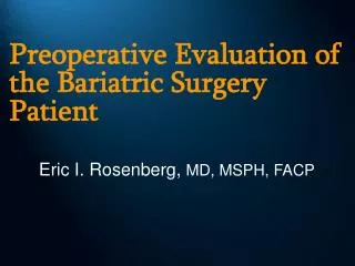
Laryngeal Height measurement Laryngeal height (cm)
Laryngeal Height measurement Reflects the degree of hyperinflation; marked hyperinflation is present if < 4 cm
Original ArticleEndovascular vs. Open Repair of Abdominal Aortic Aneurysms in the Medicare Population Marc L. Schermerhorn, M.D., A. James O'Malley, Ph.D., Ami Jhaveri, M.D., Philip Cotterill, Ph.D., Frank Pomposelli, M.D., and Bruce E. Landon, M.D., M.B.A. N Engl J Med Volume 358(5):464-474 January 31, 2008
Study Overview • Endovascular repair is a less invasive strategy than open repair • This observational study in a large Medicare population shows that perioperative survival is superior with endovascular repair but that the survival advantage gradually wanes over 3 years • The survival advantage is more durable in older patients
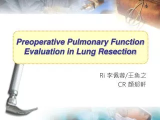
Open Repair and Endovascular Repair of an Infrarenal Abdominal Aortic Aneurysm Schermerhorn ML et al. N Engl J Med 2008;358:464-474
Baseline Characteristics of Medicare Beneficiaries Undergoing Endovascular Repair or Open Repair of Abdominal Aortic Aneurysms in the 2001-2004 Period, before and after Matching for Propensity Score Schermerhorn ML et al. N Engl J Med 2008;358:464-474
Perioperative Outcomes after Endovascular Repair or Open Repair Schermerhorn ML et al. N Engl J Med 2008; 358:464-474
Survival of Patients Undergoing Endovascular Repair or Open Repair of Abdominal Aortic Aneurysms, Overall and According to Age Schermerhorn ML et al. N Engl J Med 2008;358:464-474