Download
1 / 54
570 likes | 1.03k Views
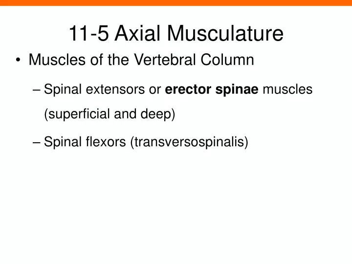
11-5 Axial Musculature. Muscles of the Vertebral Column Spinal extensors or erector spinae muscles (superficial and deep) Spinal flexors (transversospinalis) . 11-5 Axial Musculature. Muscles of the Vertebral Column Deep Spinal Extensors Semispinalis group Multifidus muscle

E N D
11-5 Axial Musculature Muscles of the Vertebral Column Spinal extensors or erector spinae muscles (superficial and deep) Spinal flexors (transversospinalis)
11-5 Axial Musculature Muscles of the Vertebral Column Deep Spinal Extensors Semispinalis group Multifidus muscle Interspinalis muscles Intertransversarii muscles Rotatores muscles
Figure 11-10 Muscles of the Vertebral Column Erector Spinae, Deep Layer Erector Spinae, Superficial Layer Splenius capitis Semispinalis Group Spinalis, Longissimus, and Iliocostalis Group Semispinalis capitis Semispinalis cervicis Longissimus capitis Spinalis cervicis Semispinalis thoracis Longissimus cervicis Iliocostalis cervicis Iliocostalis thoracis Multifidus Longissimus thoracis Spinalis thoracis Iliocostalis lumborum
11-5 Axial Musculature Muscles of the Vertebral Column Superficial Spinal Extensors Spinalis group Longissimus group Iliocostalis group
11-5 Axial Musculature Muscles of the Vertebral Column Spinal Flexors Neck Longus capitis and longus colli Rotate and flex the neck Lumbar Quadratus lumborum muscles Flex spine and depress ribs
Figure 11-10 Muscles of the Vertebral Column Erector Spinae, Deep Layer Intervertebral Muscles, Posterior View Spinal Flexors Quadratus lumborum Rotatores Intertransversarii Flexors of the Anterior Cervical and Thoracic Spine Longus capitis Spinous process of vertebra Thoracodorsal fascia Posterior view Longus colli Interspinales Transverse process of vertebra
11-5 Axial Musculature Oblique and Rectus Muscles Lie within the body wall Oblique muscles Compress underlying structures Rotate vertebral column Rectus muscles Flex vertebral column Oppose erector spinae
11-5 Axial Musculature Oblique Muscles Cervical region Scalene muscles Flex the neck Thoracic region Intercostal muscles (external and internal) Respiratory movements of ribs Transversus thoracis Cross inner surface of ribs
11-5 Axial Musculature Oblique Muscles Abdominopelvic region (same pattern as thoracic) External oblique muscles Internal oblique muscles Transversus abdominis
11-5 Axial Musculature Rectus Muscles Rectus abdominis Between xiphoid process and pubic symphysis Divided longitudinally by linea alba Divided transversely by tendinous inscriptions
11-5 Axial Musculature Rectus Muscles Diaphragmatic muscle or diaphragm Divides thoracic and abdominal cavities Performs respiration
Figure 11-11a Oblique and Rectus Muscles and the Diaphragm Scalenes Anterior Middle Posterior Anterior view, cervical region
Figure 11-11b Oblique and Rectus Muscles and the Diaphragm Serratus anterior Internal intercostal External oblique External intercostal Tendinous inscription External oblique (cut) Internal oblique Cut edge of rectus sheath Linea alba Rectus abdominis Anterior view
Figure 11-11c Oblique and Rectus Muscles and the Diaphragm Rectus abdominis Xiphoid process External oblique Costal cartilages Inferior vena cava External intercostal T10 Internal intercostal Central tendon of diaphragm Esophagus Serratus anterior Diaphragm Thoracic aorta Spinal cord Erector spinae group Superior view at the level of the diaphragm
Figure 11-11d Oblique and Rectus Muscles and the Diaphragm Rectus abdominis Rectus sheath Linea alba External oblique L3 Transversus abdominis Quadratus lumborum Internal oblique Thoracolumbar fascia Horizontal section view at the level of the umbilicus
11-5 Axial Musculature Muscles of the Pelvic Floor Functions of pelvic floor muscles Support organs of pelvic cavity Flex sacrum and coccyx Control movement of materials through urethra and anus
11-5 Axial Musculature Muscles of the Pelvic Floor Perineum Muscular sheet forming the pelvic floor, divided into: Anterior urogenital triangle Posterior anal triangle
11-5 Axial Musculature Perineum Urogenital diaphragm Deep muscular layer between pubic bones Supports the pelvic floor And muscles of the urethra Superficial muscles of the urogenital triangle Support external genitalia
11-5 Axial Musculature Muscles of the Pelvic Floor Perineum Pelvic diaphragm Deep muscular layer extending to pubis Supports anal triangle
Figure 11-12a Muscles of the Pelvic Floor Superficial Dissections Ischiocavernosus Bulbospongiosus Vagina Superficial transverse perineal Anus Gluteus maximus Female
Figure 11-12a Muscles of the Pelvic Floor Deep Dissections Urethra Urogenital Diaphragm External urethral sphincter Deep transverse perineal Central tendon of perineum Pelvic Diaphragm Pubococcygeus Levator ani Iliococcygeus External anal sphincter Coccygeus Sacrotuberous ligament Female
Figure 11-12b Muscles of the Pelvic Floor Superficial Dissections Testis Urethra (connecting segment removed) Ischiocavernosus Bulbospongiosus Superficial transverse perineal Anus Gluteus maximus Male
Figure 11-12b Muscles of the Pelvic Floor Deep Dissections UROGENITAL TRIANGLE OF PERINEUM Urogenital Diaphragm External urethral sphincter Deep transverse perineal Central tendon of perineum Pelvic Diaphragm Pubococcygeus Levator ani Iliococcygeus External anal sphincter Coccygeus Sacrotuberous ligament ANAL TRIANGLE Male
11-6 Appendicular Musculature Appendicular Muscles Position and stabilize pectoral and pelvic girdles Move upper and lower limbs Two divisions of appendicular muscles Muscles of the shoulders and upper limbs Muscles of the pelvis and lower limbs
Figure 11-13a An Overview of the Appendicular Muscles of the Trunk Superficial Dissection Axial Muscles Platysma Appendicular Muscles Deltoid Pectoralis major Latissimus dorsi Serratus anterior Anterior view ATLAS: Plates 25; 39b
Figure 11-13a An Overview of the Appendicular Muscles of the Trunk Deep Dissection Axial Muscles Sternocleidomastoid Appendicular Muscles Trapezius Subclavius Deltoid (cut and reflected) Pectoralis minor Subscapularis Pectoralis major (cut and reflected) Coracobrachialis Biceps brachii Teres major Serratus anterior Anterior view ATLAS: Plates 25; 39b
Figure 11-13a An Overview of the Appendicular Muscles of the Trunk Superficial Dissection Axial Muscles External oblique Rectus sheath Superficial inguinal ring Appendicular Muscles Tensor fasciae latae Sartorius Rectus femoris Anterior view ATLAS: Plates 25; 39b
Figure 11-13a An Overview of the Appendicular Muscles of the Trunk Deep Dissection Axial Muscles External intercostal Internal intercostal Internal oblique (cut) External oblique (cut and reflected) Rectus abdominis Transversus abdominis Appendicular Muscles Gluteus medius Iliopsoas Pectineus Adductor longus Gracilis Anterior view ATLAS: Plates 25; 39b
Figure 11-13b An Overview of the Appendicular Muscles of the Trunk Superficial Dissection Axial Muscles Sternocleidomastoid Appendicular Muscles Trapezius Deltoid Infraspinatus Teres minor Teres major Triceps brachii Posterior view ATLAS: Plate 40a,b
Figure 11-13b An Overview of the Appendicular Muscles of the Trunk Deep Dissection Axial Muscles Semispinalis capitis Splenius capitis Appendicular Muscles Levator scapulae Supraspinatus Rhomboid minor Rhomboid major Posterior view ATLAS: Plate 40a,b
Figure 11-13b An Overview of the Appendicular Muscles of the Trunk Superficial Dissection Appendicular Muscles Latissimus dorsi Thoracolumbar fascia Iliac crest Gluteus medius Gluteus maximus Posterior view ATLAS: Plate 40a,b
Figure 11-13b An Overview of the Appendicular Muscles of the Trunk Deep Dissection Axial Muscles Erector spinae muscle group External oblique Posterior view ATLAS: Plate 40a,b
11-6 Appendicular Musculature Muscles of the Shoulders and Upper Limbs Four groups Muscles that position the pectoral girdle Muscles that move the arm Muscles that move the forearm and hand Muscles that move the hand and fingers
11-6 Appendicular Musculature Muscles That Position the Pectoral Girdle Trapezius Superficial Covers back and neck to base of skull Inserts on clavicles and scapular spines
Figure 11-14a Muscles That Position the Pectoral Girdle Muscles That Position the Pectoral Girdle Trapezius Levator scapulae Subclavius Pectoralis minor Pectoralis major (cut and reflected) Internal intercostals External intercostals T12 Anterior view ATLAS: Plates 39a-d; 40a–b
Figure 11-14b Muscles That Position the Pectoral Girdle Superficial Dissection Muscles That Position the Pectoral Girdle Trapezius Posterior view ATLAS: Plates 27b; 40a–b
11-6 Appendicular Musculature Muscles That Position the Pectoral Girdle Rhomboid and levator scapulae Deep to trapezius Attach to cervical and thoracic vertebrae Insert on scapular border
Figure 11-14b Muscles That Position the Pectoral Girdle Deep Dissection Muscles That Position the Pectoral Girdle Levator scapulae Rhomboid minor Rhomboid major Scapula Serratus anterior Triceps brachii T12 vertebra Posterior view ATLAS: Plates 27b; 40a–b
11-6 Appendicular Musculature Muscles That Position the Pectoral Girdle Serratus anterior On the chest Originates along ribs Inserts on anterior scapular margin
11-6 Appendicular Musculature Muscles That Position the Pectoral Girdle Subclavius Originates on ribs Inserts on clavicle Pectoralis minor Attaches to scapula
Figure 11-14a Muscles That Position the Pectoral Girdle Muscles That Position the Pectoral Girdle Trapezius Levator scapulae Subclavius Pectoralis minor Pectoralis major (cut and reflected) Internal intercostals External intercostals T12 Anterior view ATLAS: Plates 39a-d; 40a–b
Figure 11-14a Muscles That Position the Pectoral Girdle Muscles That Position the Pectoral Girdle Pectoralis minor (cut) Serratus anterior Biceps brachii, short head Biceps brachii, long head T12 Anterior view ATLAS: Plates 39a-d; 40a–b
Table 11-11 Muscles That Position the Pectoral Girdle (Figures 11–13, 11–14)
11-6 Appendicular Musculature A&P FLIX Muscles of the Pectoral Girdle (a) A&P FLIX Muscles of the Pectoral Girdle (b) A&P FLIX Muscles of the Pectoral Girdle (c)