Download
1 / 14
140 likes | 147 Views
SEM Imaging. - Magnification and Deflection System - Focussing (contamination dots) - Imaging. To be good in e-beam lithography. means to be good in SEM imaging. Motivation. Tip Geometry & Crossover. a) LaB 6 gun. b) thermal field emission.

E N D
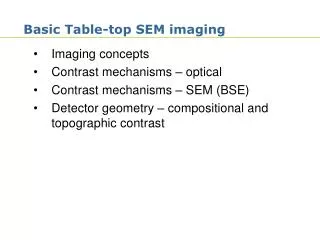
SEM Imaging - Magnification and Deflection System - Focussing (contamination dots) - Imaging
To be good in e-beam lithography means to be good in SEM imaging. Motivation
Tip Geometry & Crossover a) LaB6 gun b) thermal field emission (Gersley, J. Appl. Phys. 65 (3), 914 (1989))
source type brightness source energy vacuum [A/cm²/sr] size spread [eV] [torr] tungsten ~10 25µm 2 - 3 10 5 - 6 thermionic LaB thermionic ~10 10µm 2 - 3 10 6 - 8 6 thermal FE ~10 20nm 0.9 10 8 - 9 (Schottky) cold FE ~10 5nm 0.22 10 9 - 10 Filament Types SPIE HANDBOOK OF MICROLITHOGRAPHY, MICROMACHINING AND MICROFABRICATION Volume 1: Microlithography, Chapter 2.2
Beam deflection(E-static/magnetic) Either magnetic or electrostatic fields can be used to focus electrons just as glass lenses are used to focus rays of light. Electro-static Electro-magnetic F = q · (E+ v B) EM lenses more simple Fast deflection
Beam deflection(E-static/magnetic) Electron lenses have very poor performance (spherical and chromatic aberrations) compared to light lenses; thus electrons must be kept very close to the axis - small Spherical aberration Chromatic aberration
Beam Blanker Beam blanker off Beam blanker on Filament Anode +250V GND Beam blanker Aperture
Focussing - Contamination Dots top view side view (Images taken during the acceptance test of Raith 200 at the University of Neuchâtel (Switzerland) 4/98)
Focussing = WD and Stigmation (Image taken at KTH Stockholm (Sweden), see http://www.nanophys.kth.se/nanophys/facilities/nfl/sem-ebeam.htm)
Influence of Acceleration Voltage Low (short penetration depth) High (large penetration depth) + Clear surface structures + Less damage + Less charge up + Less edge effect – Lower resolution + Higher resolution – Unclear surface structures – More edge effects – More sample damage (heating) (A guide to Scanning Microscope Observation, Jeol web page 1999)
Influence of Beam Current-Aperture Low (small aperture) High (large aperture) + Higher resolution + Less damage (heating) + Larger depth of focus – Grainy image + Smooth image + Good signal-to-noise – Deteriorated resolution – More damage (heating) – Lower resolution – Smaller depth of focus (A guide to Scanning Microscope Observation, Jeol web page 1999)
Influence of Working Distance Small Large + Higher resolution – Smaller depth of focus + Larger depth of focus – Lower resolution (A guide to Scanning Microscope Observation, Jeol web page 1999)
Important rule for SEM imaging Take always the low magnification images first!!!