Download

1 / 86
890 likes | 915 Views
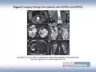
ADPKD. Kidney cysts. Cysts are abnormal blisters that may contain fluid or other matter. Kidney cysts are classified by : Cause – inherited, acquired kidney disease or advancing age Features – like the number of cysts (one or more) and whether the cysts are simple or complicated .

E N D
Kidney cysts • Cysts are abnormal blisters that may contain fluid or other matter. Kidney cysts are classified by: • Cause– inherited, acquired kidney disease or advancing age • Features – like the number of cysts (one or more) and whether the cysts are simple or complicated. • Location – outer (cortex) or inner (medulla) part of the kidney
Bosniak Classification of Renal Cysts Class I: Simple benign cysts with a well-defined homogeneous mass, a thin wall. These lesions do not enhance. Class II: Minimally complicated cysts with smooth thin internal deputations, thin peripheral rim of calcification in its wall or septa. These lesions do not show enhancement Class IIF: Minimally complicated cysts,? hyper dense, contain more calcium in the wall & may have thicker internal deputations. Follow up scanning is needed. Class III: More complicated cystic structures with irregular thickened septa, wall thickening, solid non-enhancing mural nodules, or irregular calcifications. Surgical exploration. Class IV: Malignant cyst, with non-uniform wall thickening, have irregular margins, and/or contain solid components that enhance on CT (Total nephrectomy is warranted).
Pathophysiology Cysts develop from renal tubule segments and most detach from the parent tubule after they grow to a few millimeters in size. Cyst development is generally attributed to: Increased proliferation of tubular epithelium Abnormalities in tubular cilia Excessive fluid secretion
Markedly enlarged polycystic kidneys from a patient with ADPKD in comparison to a normal kidney in the middle.
DEFINITION ADPKD is a multisystem disorder characterized by multiple, bilateral renal cysts associated with cysts in other organs, such as liver, pancreas, and arachnoid membranes. It is a genetic disorder mediated primarily by mutations in two different genes and is expressed in an autosomal dominantpattern, with variable expression. Approximately 5% of patients who initiate dialysis annually in the United States.
PKD Genetics Incidence • Autosomal Dominant 1:500-1,000 live births • Autosomal Recessive 1:6,000-40,000 live births
Genes in PKD GeneProtein ADPKD1 Polycystin 1 ADPKD2 Polycystin 2 ARPKD Fibrocystin /polyductin
Median Age of ESRD PKD1 ~ 53 years PKD2 ~ 73 years Presence of at least one affected family member who developed ESRD </= 55 years was highly predictive of PKD1 mutation (PPV 100%, sensitivity 72%) If family member developed ESRD at age >/= 70 years, then highly predictive of PKD 2 mutations (PPV 100%, Sensitivity 74%) Family History of Severity of Renal Disease Predicts Mutated Gene in ADPKD Barau et al JASN20: 1833, 2009
Etiology and Pathogenesis The polycystic kidney disease (PKD) proteins now known as polycystin 1 (PC1)and polycystin 2 (PC2) play a critical role in the normal function of the primary cilium that is essential to maintaining the differentiated phenotype of tubular epithelium. Disordered function of polycystins is the basis for cyst formation in PKD by permitting a less differentiated tubular epithelial phenotype.
Fluid Secretion into Cysts Increased cAMP promotes cyst growth and overall enlargement increased transepithelial secretion of chloride through apical CFTR channels Cystogenesis
Diagnosis Imaging tests the gold standard At present, asymptomatic screen not recommended Ultrasound: false negative rate 16-18% before age 30 CT, MR: probably more sensitive
Genetic Testing DNA screen of polycystin 1 and polycystin 2 available Up to 90% detection rate – better with affected family members Expensive
Differential Diagnosis • Syndromes Mimicking ADPKD • Von Hippel-Lindau • Tuberous Sclerosis • ARPKD • Simple Renal Cysts • Acquired Cystic Disease of Renal Failure • Medullary Cystic Disease / Nephronopthisis
Clinical Manifestations Renal and extrarenalmanifestations of ADPKD have been described that cause significant complications.
Renal Complications 1. Hypertension 60-100% 2. Gross hematuria 50% 3. Infection common 4. Nephrolithiasis 20-25% 5. Renal failure 50% by age 60 (PKD1)
Mechanisms of Hypertensionin ADPKD Normal kidney ADPKD kidney
Pathogenetic role of RAAS in ADPKD Schrier JASN 20:1888-1893, 2009
Hypertension in PKD Control most important to prevent progression ACE inhibitors, ARBs theoretically better Blood pressure goal not established (should be to less than 130/80 mmHg) BP goal of less than 120/80 mmHg may provide cardiovascular benefit among ADPKD patients with left ventricular hypertrophy
Nephrolithiasis in ADPKD ~20-36% of patients Uric acid (UA) (~50%); Ca Ox (~47%) Predisposing factors: hypocitraturia, hyperoxaluria, hypercalciuria, urinary stasis from expanding cysts, Low urine pH Rx: K-citrate, Alkalinisationof urine Lithotripsy can be performed safely, but residual fragments in ~50% of patients
Hematuria in ADPKD Cyst hemorrhage occurs in~60% of individuals Gross or microscopic hematuria if cyst connects to collecting system Susceptible to minor trauma with resultant hemorrhage Patients with recurrent episodes of gross hematuria have the largest kidneys and progress more quickly to kidney failure Conservative management with hydration, bed rest, and appropriate use of analgesics Rarely, massive bleeding may require transfusion, kidney embolization or nephrectomy
Kidney Infection in ADPKD 30 to 50% (more common in women) Cyst infections ~ 0.01 episode/patient/year (10% of causes that led to hospitalization) Fever and Flank pain are the presenting symptoms Urine culture may be negative in cyst infection, as cysts frequently don’t communicate with the collecting system. (E coli ~ 75% of cases ) +ve urine culture ~39%, +ve blood culture ~24% in renal cysts infection
Infected cysts in PKD Localization of infected cysts is difficult • Labeled WBC or gallium scan (positive in ~50% of cases) • CT&MRI with contrast (good for R/O renal or perinephric abscesses) • PET scan • Ultrasound, CT scan, MRI, and PET scan yielded positive results in 6, 18, 40, and 100%, respectively for infected cysts Sallée et. al. Clin J Am SocNephrol 4: 1183-1189, 2009
Antibiotics in PKD Some drugs do not penetrate cysts well Fluoroquinolones, Tmp-Sulfa, chloramphenicol best for cyst penetration Percutaneous or operative drainage is rarely needed; only refractory infection Complicated upper tract: cyst penetratingantibiotics 3-4 weeks
Hepatic and pancreatic cysts Asymptomatic in many patients, but can expand and cause pain and infection; rarely massive PLD Cardiac valvular abnormalities Mitral valve prolapse, tricuspid and aortic regurgitation Intracranial aneurysms Found in approximately 5% of patients with no family history and about 22% of patients with family history of ICA or SAH Seminal vesicle cysts Found in ~39-60% of men; undefined risk of infertility Principal Extra renal Manifestations
Liver/GI Complications 1. Liver cysts (94% > 35) - asymptomatic up to 80% - symptomatic uncommon (W:M 10:1) 2. Pancreatic cysts ~10% 3. Intestinal diverticuli ~80% pts with ESRD 4. Hernias ~10%
Liver Cysts: Sx and Infection With marked hepatomegaly: Heaviness, dull ache, Mechanical low back pain, Early satiety If fever persists 1-2 wks after antibiotics in infected cysts, drainage frequently needed Hepatic cyst infection more serious than renal cyst infection. Do not delay drainage esp >5cm CA 19-9: Marker for Hepatic Cyst Infection? Rise (active infection)/ fall (improvement) Kanaan et al AJKD March, 2010
Vascular manifestations of ADPKD. • A, Gross specimen demonstrating bilateral aneurysms of the middle cerebral arteries. • 90% of aneurysms in anterior circulation • 10% of aneurysms in posterior circulation (greater risk of rupture) • B, Gross specimen demonstrating a thoracic aortic dissection extending into the abdominal aorta in a patient with ADPKD.
Cerebral Aneurysm in ADPKD • No family history – 6% prevalence • Family history – 21% prevalence • Clinical Symptoms (Ruptured) – pain, stiff neck, coma; >50% mortality • The mean age of rupture of intracranial aneurysms is lower in individuals with ADPKD than in the general population (39 years vs 51 years)
Risk of ICA Rupture in ADPKD %YrAneurysm Size 0.05 <10 mm 1.0 10-24 mm 6.0 >24 mm 0.05% per year <10mm. No personal or family hx of SAH 0.5% per year<10 mm. With personal or family hx of SAH Betz KI 63:2003 Gibbs KI 64:2004
Risk Factors for ICA Rupture • Most aneurysms have a very low risk of rupture and occurs without a family history • With 2 PKD relatives with SAH the RR= 2.15 • F>M and ICA>8mm • Pack years of smoking • HTN > 10 years • Torres 2009
F/U of ICA in ADPKD <7 mm Observation 7-12 mm Risk assessment >12 mm Intervene Follow-up with CTA or MRA annually for two to three years, and every two to five years thereafter if the aneurysm is clinically and radiographically stable. It is not unreasonable to reimage newly detected small aneurysms at six months.