Download
1 / 43
430 likes | 454 Views
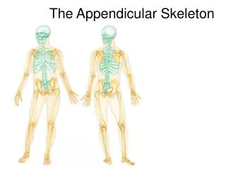
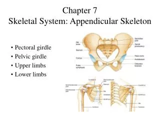
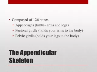
Essentials of Human Anatomy The Skeletal System 3 Appendicular Skeleton. Dr Fadel Naim Ass. Prof. Faculty of Medicine IUG. 1. The Appendicular Skeleton. Pectoral girdle Attaches the upper limbs to the trunk Pelvic girdle Attaches the lower limbs to the trunk

E N D
Essentials of Human AnatomyThe Skeletal System 3Appendicular Skeleton Dr Fadel Naim Ass. Prof. Faculty of Medicine IUG 1
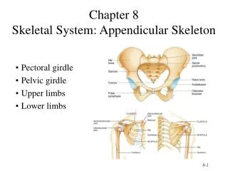
The Appendicular Skeleton • Pectoral girdle • Attaches the upper limbs to the trunk • Pelvic girdle • Attaches the lower limbs to the trunk • Upper and lower limbs differ in function • Share the same structural plan
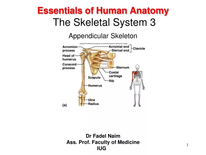
Appendicular Skeleton • Upper extremity • Consists of the bones of the shoulder girdle, upper arm, lower arm, wrist, and hand • Shoulder girdle • Made up of scapula and clavicle • Clavicle forms only bony joint with trunk, the sternoclavicular joint • At its distal end, clavicle articulates with the acromion process of the scapula
Pectoral Girdle • shoulder girdle • clavicles • scapulae • supports upper limbs
Clavicles • articulate with manubrium • articulate with scapulae (acromion process)
Scapulae • spine • supraspinous fossa • infraspinous fossa • acromion process • coracoid process • glenoid cavity
The Upper Limb • 30 bones form each upper limb • Grouped into bones of the • Arm • Forearm • Hand
Upper Limb • Humerus • Radius • Ulna • Carpals • Metacarpals • Phalanges
Humerus • The long bone of the upper arm • Articulates proximally with the glenoid fossa of the scapula and distally with the radius and ulna
Humerus • head • greater tubercle • lesser tubercle • anatomical neck • surgical neck • deltoid tuberosity • capitulum • trochlea • coronoid fossa • olecranon fossa
Radius • Long bone found on thumb side of forearm • Articulates proximally with capitulum of humerus and radial notch of ulna • articulates distally with scaphoid and lunate carpals and with head of ulna
Radius • lateral forearm bone • head • radial tuberosity • styloid process
Ulna • Long bone found on little finger side of forearm • Articulates proximally with humerus and radius and distally with a fibrocartilaginous disk
Ulna • medial forearm bone • trochlear notch • olecranon process • coronoid process • styloid process
Wrist and Hand • Carpals (16) • trapezium • trapezoid • capitate • scaphoid • pisiform • triquetrum • hamate • lunate • Metacarpals (10) • Phalanges (28) • proximal phalanx • middle phalanx • distal phalanx
Appendicular Skeleton • Lower extremity Consists of the bones of • Hip • Thigh • lower leg • Ankle • Foot
Pelvis • The adult pelvis is composed of four bones: • the sacrum, the coccyx, and the right and left ossa coxae. • Protects and supports the viscera in the inferior part of the ventral body cavity. • Pelvic girdle refers to the left and right ossa coxae only.
Os Coxae • Commonly referred to as the “hip bone” or innominate bone. • Each is formed from three separate bones: • the ilium • the ischium • the pubis • Each articulates posteriorly with the sacrum at the sacroiliac joint.
Pelvic Girdle • Coxae (2) • supports trunk of body • protects viscera
Coxae • hip bones • acetabulum • ilium • iliac crest • iliac spines • greater sciatic notch • ischium • ischial spines • lesser sciatic notch • ischial tuberosity • pubis • obturator foramen • symphysis pubis • pubic arch
Greater and Lesser Pelvis • Lesser Pelvis • sacrum and coccyx posteriorly • lower ilium, ischium, and pubis bones laterally and anteriorly • Greater Pelvis • lumbar vertebrae posteriorly • iliac bones laterally • abdominal wall anteriorly
Male and Female Pelvis • Female • iliac bones more flared • broader hips • pubic arch angle greater • more distance between ischial spines and ischial tuberosities • sacral curvature shorter and flatter • lighter bones
Lower Limb • Femur • Patella • Tibia • Fibula • Tarsals • Metatarsals • Phalanges
Femur • longest bone of body • head • fovea capitis • neck • greater trochanter • lesser trochanter • linea aspera • condyles • epicondyles
Patella • kneecap • anterior surface of knee • flat sesamoid bone located in a tendon
Tibia • shin bone • medial to fibula • condyles • tibial tuberosity • anterior crest • medial malleolus
Fibula • lateral to tibia • long, slender • head • lateral malleolus • does not bear any body weight
Ankle and Foot • Tarsals (14) • calcaneus • talus • navicular • cuboid • lateral cuneiform • intermediate cuneiform • medial cuneiform • Metatarsals (10) • Phalanges (28) • proximal • middle • distal
Arches of the Foot • The sole of the foot does not rest flat on the ground. • Helps it support the weight of the body. • Ensures that the blood vessels and nerves on the sole of the foot are not pinched when standing.
Arches of the Foot • Medial longitudinal arch extends from the heel to the big toe. • Lateral longitudinal arch is not as high as the medial longitudinal arch. • Transverse arch runs perpendicular to the longitudinal arches.
Hallux valgus • A lateral deviation of the great toe at the metatarsophalangeal joint • Its incidence is greater in women than in men • Associated with badly fitting shoes. • Often accompanied by the presence of a short first metatarsal bone. • Once the deformity is established, it is progressively worsened by the pull of the flexor hallucis longus and extensor hallucis longus muscles.
Hallux rigidus • Osteoarthritic changes in the metatarsophalangeal joint, which then becomes stiff and painful
Pes planus (flat foot) • A condition in which the medial longitudinal arch is depressed or collapsed. • As a result, the forefoot is displaced laterally • The head of the talus is no longer supported • The causes of flat foot are both congenital and acquired.