Download
1 / 7

E N D
Chap. 15 Problem 2 Signaling systems are classified based on the distance over which they act. Endocrine signaling acts over long distances within the organism. Paracrine signaling acts over a very short distances, for example between neighboring cells. In autocrine signaling, cells release ligands that bind to their own surface receptors, modulating activity. Because growth hormone is synthesized by the pituitary gland in the brain and travels to the liver by the blood, growth hormone signals via an endocrine mechanism.
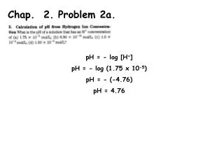
Chap. 15 Problem 3 Receptor 2 has greater affinity for the ligand than Receptor 1 because the Kd for Receptor 2 (10-9 M) is lower than the Kd for Receptor 1 (10-7 M). The fraction of receptors that are bound to the ligand when its concentration is 10-8 M can be calculated using the rearranged form of the Kd equation, [R]/[LR] = Kd/[L]. For Receptor 1, [R]/[LR] = (10-7 M)/(10-8 M) = 10/1 at this concentration of ligand. The receptor therefore is only about 10% saturated with ligand. For Receptor 2, [R]/[LR] = (10-9 M)/(10-8 M) = 1/10 at this concentration of ligand. The receptor therefore is about 90% saturated with ligand.
Chap. 15 Problem 6 The G protein cycle of activity in hormone-stimulated G protein coupled receptor (GPCR) regulation of effector proteins is summarized in Fig. 15.17 (next slide). The trimeric G protein complex is tethered to the inner leaflet of the cytoplasmic membrane via lipid anchors attached to the Ga and Gg subunits. The trimeric GDP-bound form of the G protein is inactive in signaling. The binding of a hormone to the GPCR triggers a conformational change in the receptor (Step 1) which promotes its binding to the trimeric G protein (Step 2). Binding to the activated GPCR triggers exchange of GTP for GDP and activation of the Ga subunit which dissociates from Gßg (Steps 3 & 4). Ga-GTP then binds to the effector protein regulating its activity (Step 5). In time (often < 1 min), GTP is hydrolyzed to GDP and Ga becomes inactive (Step 6). It then recombines with Gßg. If a mutation increased the GTPase activity of the G subunit, then the subunit would be active for a shorter time than normal. The activation of the downstream effector would therefore be reduced.
Chap. 15 Problem 10 (modified) channels via G proteins. Describe the rhodopsin signal transduction pathway. The rhodopsin signal transduction pathway is shown in Fig. 15.23. Light absorption by rhodopsin triggers GTP/GDP exchange on the transducin (Gat subunit, and dissociation of this trimeric G protein. Gat-GTP binds to and activates a cGMP phosphodiesterase, reducing intracellular cGMP level. This indirectly results in the closing of non-selective Na+/Ca2+ ion channels in the cytoplasmic membrane and hyperpolarization of the membrane potential. This results in decreased release of neurotransmitter from the cell.
Chap. 15 Problem 12 In the liver, muscle, and adipose tissue, epinephrine raises cAMP levels through Gas-GTP activation of adenylyl cyclase. The key target of cAMP is protein kinase A (PKA) (Fig. 15.29a). Through this point in the epinephrine-cAMP pathway, the steps of signal transduction are the same in all three tissues. However, each tissue differs in the downstream targets of PKA, resulting in cell type-specific responses.
Chap. 15 Problem 15 Phospholipase C (PLC) cleaves the membrane lipid, phosphatidylinositol 4,5-bisphosphate (PIP2) to the second messengers, inositol 1,4,5-trisphosphate (IP3) and diacylglycerol (DAG) (Fig. 15.35). The steps downstream of PLC are illustrated in Fig. 15.36a. IP3 diffuses from the cytoplasmic membrane to the ER where it binds to and triggers the opening of IP3-gatedCa2+channels. The rise in cytoplasmic [Ca2+] activates calmodulin which in turn activates certain cytoplasmic kinases, such as glycogen phosphorylase kinase. DAG binds to and activates the kinase known as protein kinase C (PKC). Cells replenish ER calcium via ER membrane Ca2+-ATPase pumps that transport calcium back to the ER lumen. These pumps also transport cytoplasmic calcium outside of the cell. Extracellular calcium is transported back into the cytoplasm by activation of store-operated calcium channels in the cytoplasmic membrane. This calcium ultimately is transported back to the ER lumen by the ER membrane Ca2+-ATPase pumps (not shown).