Download
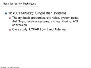
1 / 128
1.3k likes | 1.65k Views
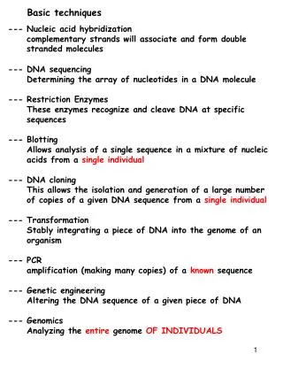
Basic techniques. --- Nucleic acid hybridization complementary strands will associate and form double stranded molecules --- Restriction Enzymes These enzymes recognize and cleave DNA at specific sequences --- Blotting Allows analysis of a single sequence in a mixture

E N D
Basic techniques --- Nucleic acid hybridization complementary strands will associate and form double stranded molecules --- Restriction Enzymes These enzymes recognize and cleave DNA at specific sequences --- Blotting Allows analysis of a single sequence in a mixture --- DNA cloning This allows the isolation and generation of a large number of copies of a given DNA sequence --- Transformation Stably integrating a piece of DNA into the genome of an organism --- DNA sequencing Determining the array of nucleotides in a DNA molecule --- PCR amplification of known sequence --- Genetic engineering Altering the DNA sequence of a given piece of DNA --- Genomics Analyzing changes in an entire genome
Nucleic acid hybridization Complementary strands of DNA or RNA will specifically associate DNA is heated to 100C, the hydrogen bonds linking the two strands are broken The double helix dissociates into single strands. As the solution is allowed to cool, strands with complementary sequences readily re-form double helixes. This is called Nucleic acid hybridization. 5’ AAAAAAAATTTTAAAAAAA 3’ Will associate with 3’ TTTTTTTTAAAATTTTTTT 5’ This occurs with complementary DNA/DNA, DNA/RNA, RNA/RNA
Li-Fraumeni syndrome This technique is very sensitive and specific. A single 200 nucleotide sequence when added to a solution of a million sequences will specifically hybridize with the ONE complementary sequence Usefulness Li-Fraumeni syndrome Individuals in a family have a propensity to develop tumors at an early age Often these families have a deletion in the p53 gene When this family has a child, they might want to know if their child has normal p53 or not Nucleic acid hybridization provides a means to rapidly determine whether the sequence is present or not
Sequencing Genomic DNA Fragment DNA (clone) Sequence fragments Align fragments Build consensus sequence ACGCGATTCA ACGCGATTCA GCGATTCAGGTTA GATTCAGGTTA CAGGTTACCACGC ACGCGTAGCGC TAGCGCA TAGCGCATTACAC ACGCGATTCAGGTTACCACGCGTAGCGCATTACAC
Sequencing Reference Genome- Number of donor DNAs are sequenced Pieces of DNA are sequenced many times Computers are used to overlap the pieces to generate contigs Consensus sequence is reference genome Sequences of individuals will vary from the reference genome ACGCGATTCAGGTTACCACGCGTAGCGCATTACAC Reference Genome ACGCGATTCAGGTTACCACGCGTAGCGCATTACAC MISTY ACGCGGTTCAGGTTACCACGCGTAGCGCATTACAC NICK ACGCGATTCAGGTTACCACGCGTAAAACATTACAC JESSE ACGCGGTTCAGGTTACCCCGCGTAGCGCATTACAC DONNA The sequence homology between Individuals is not perfect!!! This allows us to assign a specific sequence to a specific Individual
Homology (molecular biology) Regions of the DNA (gene or non-gene) that share similar nucleotide sequence Sequence homology is a very important concept Structural homology (nucleotide sequence) implies functional homology Genes with a similar sequence are likely to function in a similar manner Variation in sequence between individuals is also very Important
Isolate DNA The method normal individual Patient Fragment DNA, Heat to denature Add radiolabeled ssDNA (p53 gene) (p53 probe) Gradually and slowly cool solution Radiolabeled p53 probe associates with DNA in normal individual If patient is deficient for p53 gene Radiolabeled p53 probe is unable to associates with DNA in patient Add enzyme (nuclease) that specifically degrades ssDNA molecules. dsDNA remains degraded No radiolabel present in dsDNA (because p53 probe could not anneal) Radiolabel present in dsDNA
Restriction Enzymes What are Restriction enzymes What are restriction enzyme sites in DNA How do we map Restriction enzyme sites in DNA How do we use restriction enzymes to clone pieces of DNA How do we use restriction enzyme sites/maps to study individuals
Restriction Enzymes Enzymes which Recognize a SPECIFIC DNA sequence BIND that sequence and CUT The DNA at that specific sequence SmaI is a Restriction enzyme | 5’ AAAACCCGGGAAAA3’ 3’ TTTTGGGCCCTTTT5’ | This sequence is symmetrical. If one rotates it about the axis It reads the same EcoRI is another Restriction enzyme | 5’ AAAAGAATTCAAAA3’ 3’ TTTTCTTAAGTTTT5’ | Some restriction enzymes recognize a specific sequence that is 4 bp long Some restriction enzymes recognize a specific sequence that is 6 bp long Some restriction enzymes recognize a specific sequence that is 8 bp long
BamHI Restriction enzymes Restriction enzyme digestion of DNA (linear genomic double stranded DNA) OR Restriction enzyme digestion of bacterial plasmid DNA (small double stranded circular DNA) No digestion of RNA No digestion of single stranded DNA
Linear/Circular DNA No digestion of RNA No digestion of single stranded DNA A linear DNA molecule with ONE SmaI site will be cut into two fragments A circular DNA molecule with ONE SmaI site will generate one DNA fragment
Blunt Vs Sticky Blunt ends Sticky ends After digestion of DNA by a restriction enzyme the DNA ends are either blunt or sticky
Restriction sites SmaI- BLUNT ENDS 5’AAAAAAAAAAGGGGGGGGTTTTTTTCCCGGGAAAAAAAAGGGGGGGGTTTTTT3’ 3’TTTTTTTTTTCCCCCCCCAAAAAAAGGGCCCTTTTTTTTCCCCCCCCAAAAAA5’ 5’AAAAAAAAAAGGGGGGGGTTTTTTTCCC GGGAAAAAAAAGGGGGGGGTTTTTT3’ 3’TTTTTTTTTTCCCCCCCCAAAAAAAGGG CCCTTTTTTTTCCCCCCCCAAAAAA5’ EcoRI is another commonly used restriction enzyme 5’AAAAAAAAAAGGGGGGGGTTTTTTTGAATTCAAAAAAAAGGGGGGGGTTTTTT3’ 3’TTTTTTTTTTCCCCCCCCAAAAAAACTTAAGTTTTTTTTCCCCCCCCAAAAAA5’ 5’AAAAAAAAAAGGGGGGGGTTTTTTTG AATTCAAAAAAAAGGGGGGGGTTTTTT3’ 3’TTTTTTTTTTCCCCCCCCAAAAAAACTTAA GTTTTTTTTCCCCCCCCAAAAAA5’ Unlike SmaI which produces a blunt end, EcoRI produces sticky or cohesive ends (SINGLE STRANDED) These cohesive ends facilitate formation of recombinant DNA molecules
5’AAAAAAAAAAGGGGGGGGTTTTTTTGAATTCAAAAAAAAGGGGGGGGTTTTTT3’5’AAAAAAAAAAGGGGGGGGTTTTTTTGAATTCAAAAAAAAGGGGGGGGTTTTTT3’ 3’TTTTTTTTTTCCCCCCCCAAAAAAACTTAAGTTTTTTTTCCCCCCCCAAAAAA5’ 5’AAAAAAAAAAGGGGGGGGTTTTTTTG AATTCAAAAAAAAGGGGGGGGTTTTTT3’ 3’TTTTTTTTTTCCCCCCCCAAAAAAACTTAA GTTTTTTTTCCCCCCCCAAAAAA5’ 5’AAAAAAAAAAGGGGTTTTTTTG AATTCACGTACGTACGTACGTACGTACGTG AATTCAAAAAAAAGGGGGGGGTTTTTT3’ 3’TTTTTTTTTACCCCAAAAAAACTTAA GTGCATGCATGCATGCATGCATGCACTTAA GTTTTTTTTCCCCCCCCAAAAAA5’ 5’AAAAAAAAAAGGGGTTTTTTTGAATTCACGTACGTACGTACGTACGTACGTGAATTCAAAAAAAAGGGGGGGGTTTTTT3’ 3’TTTTTTTTTACCCCAAAAAAACTTAAGTGCATGCATGCATGCATGCATGCACTTAAGTTTTTTTTCCCCCCCCAAAAAA5’
Complementary sticky ends AATTCAAAAAAAAGGGGGGGGTTT3’ GTTTTTTTTCCCCCCCCAAA5’ AAAAAAGGGGGGGGTTTTTTTG TTTTTTCCCCCCCCAAAAAAACTTAA AAAAAAGGGGGGGGTTTTTTTG TTTTTTCCCCCCCCAAAAAAACTTAA AATTCAAAAAAAAGGGGGGGGTTT3’ GTTTTTTTTCCCCCCCCAAA5’ GGCCCAAAAAAAAGGGGGGGGTTT3’ GTTTTTTTTCCCCCCCCAAA5’ AAAAAAGGGGGGGGTTTTTTTG TTTTTTCCCCCCCCAAAAAAACTTAA
Enzyme compatibility SmaI AAACCCGGGAAA XmaI AAACCCGGGAAA TTTGGGCCCTTT TTTGGGCCCTTT EcoRI AAAGAATTCAAA MfeI AAACAATTGAAA TTTCTTAAGTTT TTTGTTAACTTT KpnI AAAGGTACCAAA Asp718 AAAGGTACCAAA TTTCCATGGTTT TTTCCATGGTTT
Restriction maps Restriction maps are descriptions of the number, type and distances between Restriction sites on a piece of DNA. Very useful for molecular biologists. Previously we used specific genes as markers on chromosome and Map units to indicate distance between the markers. Its like using specific landmarks to identify your location along a road. Restriction sites are also used as landmarks along a piece of DNA. 11Mu 5Mu pr vg cy Vg Pr 205kb 300kb 4kb PstI SmaI EcoRI HindIII NNNNNNNNNGAATTCNNNNNNNNNNNNAAGCTTNNNNNNNNNNNNCTGCAGNNNNNNNNNNCCCGGGNNNNNN NNNNNNNNNCTTAAGNNNNNNNNNNNNTTCGAANNNNNNNNNNNNGACGTCNNNNNNNNNNGGGCCCNNNNNN AAAAAAAAAGAATTCTTTTTTTTTTTTAAGCTTCCCCCCCCCCCCCTGCAGGGGGGGGGGGCCCGGGAAAAAA TTTTTTTTTCTTAAGAAAAAAAAAAAATTCGAAGGGGGGGGGGGGGACGTCCCCCCCCCCCGGGCCCTTTTTT Restriction sites CAN serve as MARKERS ALONG the DNA. They can be used to generate a physical map of a specific DNA sequence can be created.
Restriction maps Human Genome is 1.5 billion basepairs long There are 25,000 genes (markers) Every gene is on average approximately 600,000 bp apart EcoRI sites are on average 4000 bp apart HinDIII sites are on average 4000 bp apart Etc etc There are many more Restriction enzyme sites (landmarks) on any one piece of DNA then there are genes (landmarks)
Human Chimp Gibbon Sequence Divergence The restriction map is a partial picture of the nucleotide sequence of a gene. By comparing restriction maps we can surmise differences in the sequence between species NNNNNNNNGAATTCNNNNNNNNNNNNNNNNAAGCTTNNNNNNNNNNNNNNCTGCAGNNNNNNNNNNNNNNN NNNNNNNNCTTAAGNNNNNNNNNNNNNNNNTTCGAANNNNNNNNNNNNNNGACGTCNNNNNNNNNNNNNNN GeneA Mai California me rahta hu aur UCSC me padhata hu. Mai California me rahta hu aur UCSC and UCLA me padhata hu. Mai California me rahta hu aur mai sirf UCLA me padhta hu
Deletions and additions Normal Globin gene 3 5 8 4 EcoRI EcoRI EcoRI HindIII HindIII Globin gene from a thallesimia patient 3 5 3 4 EcoRI EcoRI EcoRI HindIII HindIII With restriction maps, the relationship between a gene from two different individuals can be determined without having to actually sequence the gene from both individuals.
Very Large deletions or insertions can be studied using microscopy Small to large deletions/insertions (100 bp to several kb) can be studied using restriction maps!!
Restriction map Describing a DNA piece based on the pattern of restriction sites Restriction map of a cloned piece of DNA Linear or circular H E E B B K K S S B B How do I do this for an unknown piece of DNA?
Analogy Goal: Identify the parts for this blob and describe the spatial relationship between the parts 1 2 Break the blob apart into its separate parts Look at the shapes and numbers of the parts a c a 3 Fit the parts together c b b
Method Restriction map of piece of DNA Large amount of pure DNA (many copies of the same DNA is required) (Cloned) Take (cloned) DNA in a tube, Add restriction enzyme, Allow enzyme to cut DNA at its binding sites Run the digested DNA on a gel to resolve the DNA fragments based on size Piece the fragments together to determine the linear order of the fragments (build the map)
EcoRI/HindIII HindIII Marker EcoRI - + 1 3 5 2 EcoRI EcoRI EcoRI HindIII HindIII Gel electrophoresis Agarose gel electrophoresis The length of the DNA can be accurately determined by allowing the charged DNA to run through an agarose gel. DNA is an anion (-ve charged) and moves towards the Positive anode. The rate of migration of a DNA fragment is inversely proportional to its size. Larger the size, slower its movement. EcoRI/HindIII HindIII Marker EcoRI 7 5 4 3 2 1
12 2 6 Mapping You are given a 20 kb fragment of DNA After trying many enzymes you find that EcoRI and HindIII cut the fragment HindIII 14kb and 6kb EcoRI 12kb 6kb and 2kb Solve the map HindIII Marker EcoRI uncut H 6 14 20 14 Or its mirror image 12 6 14 6 4 They are the same 2 14 6 What about EcoRI? 1 E E How do you arrange these three fragments wrt one another
Mapping Since HindIII cut the 20kb fragment once, in which of the three EcoRI fragment does it cut? A double digest with both enzymes will provide the answer Fragments of 8kb, 6kb, 4kb and 2kb The double digest does not alter the size of the 6kb and 2kb fragments The 12kb fragment is lost. Also 8+4=12 EcoRI+HindIII HindIII Marker EcoRI 14 12 8 6 2 6 12 4 4 2 H E 6 E 2 8 4 1 Partial Map only!!!! Not enough information
New Mapping How are these fragments ordered? The HindIII single digest tells us that they must be ordered so that One side adds up to 6kb and the other side adds up to 14kb EcoRI+ HindIII HindIII Marker EcoRI 14 12 6 4 2 1
Mapping HindIII EcoRI HindIII/EcoRI 14 12 8 6 6 6 4 2 2
Mapping HindIII EcoRI HindIII/EcoRI 14 12 8 6 6 6 4 2 2 HindIII 4 8 12 & 8 6 2 12 4 8 16 & 4 6 2 12 4 8 10 & 10 6 2 12 4 8 14 & 6 6 2 12 4 8 12 & 8 6 2 12 4 8 16 & 4 6 2 12
Another linear map EcoRI+ HindIII PstI EcoRI+ PstI HindIII Marker EcoRI 14 12 8 6 4 2 1
Different Mapping example Hi Ec Hi/Ec 12 12 8 8 6 6 4 2 2 Ps Ps/Ec 13 12 7 5 2 1 Three different enzymes Hi Ec Ps
Mapping HindIII EcoRI HindIII/EcoRI 12 12 8 8 6 6 4 2 2 HindIII 12 & 8 12 & 8
Mapping HindIII EcoRI HindIII/EcoRI 12 12 8 8 6 6 4 2 2 16 12 HindIII 8 4 8 4 H 6 6 2 2 12 12 16 & 4 12 E E 4 8 H 2 6 12 12 & 8 E E H 12 & 8 E E
Mapping EcoRI PstI PstI/EcoRI 12 13 12 6 7 5 2 2 1
Mapping EcoRI PstI PstI/EcoRI 12 13 12 6 7 5 2 2 1 P Pst I 1 5 5 & 15 6 2 12 E E P 1 5 1 & 19 6 2 12 E E 2 6 12 E E P 1 5 3 & 17 2 6 12 E E P 1 5 7 & 13 2 6 12 E E 4 8 H
Final Map P 5 1 8 4 2 E E H Hi Ec Hi/Ec 12 12 8 8 6 6 4 2 2 Ps Ps/Ec 13 12 7 5 2 1
Mapping deletions Say you isolated this DNA from a region coding for GeneA, from a normal Patient and one suffering from a syndrome. The fragment was 17kb in the affected individual rather than 20kb in the normal patient You draws restriction maps for the normal patient 4 6 E 8 H E 2 You draws restriction maps for the affected individual 1 6 E 8 H E 2 There is a 3kb deletion in the 4kbHindIII/EcoRI fragment
Complex Mapping Often maps are more complex and difficult to analyze using single and double digests alone. To simplify the analyses, you can isolate each EcoRI band from the gel and then digest with HindIII 2kb+HindIII 12kb+HindIII 6kb+HindIII EcoRI+ HindIII Marker Marker HindIII Marker Marker 2kb 12kb EcoRI 6kb 14 14 14 14 12 12 12 12 6 6 6 6 4 4 4 4 2 2 2 2 1 1 1 1
Mt DNA Y chromosomes can be used to study paternal lineage mtDNA can be used to follow maternal lineage Cells contain organelles- Mitochondria are organelles that produce Energy. They contain a small 17,000 bp circular DNA. It encodes for 13 proteins in human cells and some tRNA’s Hypervariable region (150 bp) tRNA CytochromeB NADH dehydrogenase cytochromeC oxidase ATP synthase Mitochondrial DNA inheritance is not mendelian It is inherited maternally
Using DNA to study History This hypothesis was initially derived from restriction maps of mitochondrial DNA Australia Europe Asia Africa “Eve’s DNA” All humans are derived from a small African population about 170K yrs ago
Eve Geographic region DNA A Mutation generates B from A. Now you have individuals With A and B DNA in population. C B A B A D
Out of Africa C E C C F B A B A G D D C migrates to form a separate population. Additional mutations diversify DNAs in populations. Original population more diverse than newer population Compared sequences of mtDNA There are greater sequence differences among Africans than any other group (Europeans, American Indians, Asians, etc etc) The african population had the longest time to evolve variation And thus humans originated in Africa.
THE GENE PROBE!!! How do we isolate a GENE? How did we get a pure copy of the gene?
Cloning DNA A reasonable question is how did we clone a fragment of DNA Or how do we clone a gene The construction of Recombinant DNA molecules or cloning of DNA molecules Recombinant DNA is generated through cutting and pasting of DNA to produce novel sequence arrangements Restriction enzymes such as EcoRI produce staggered cuts leaving short single-stranded tails at the ends of the fragment. These “cohesive or sticky” ends allow joining of different DNA fragments | nnnGAATTCnnn nnnCTTAAGnnn | When a piece of DNA is cut with EcoRI, you get nnnG AATTCnnn nnnCTTAA Gnnn
Cloning DNA A reasonable question is how did we get the 20kb fragment of DNA in the first place? To understand the origin of the fragment we must address the issue of: The construction of Recombinant DNA molecules or cloning of DNA molecules Recombinant DNA is generated through cutting and pasting of DNA to produce novel sequence arrangements
Recombinant DNA Restriction enzymes such as EcoRI produce staggered cuts leaving short single-stranded tails at the ends of the fragment. These “cohesive or sticky” ends allow joining of different DNA fragments | GAATTC CTTAAG | When a piece of DNA is cut with EcoRI, you get AATTC-----------------G G-----------------CTTAA AATTC-----------------G G-----------------CTTAA 5’AAAAAAAAAAGGGGTTTTTTTG AATTCAAAAAAAAAAAAAAGGGGGGGGTTTTTTTG AATTCAAAAAAAAGGGGGGGGTTTTTT3’ 3’TTTTTTTTTACCCCAAAAAAACTTAA GTTTTTTTTTTTTTTCCCCCCCCAAAAAAACTTAA GTTTTTTTTCCCCCCCCAAAAAA5’