Download

1 / 28
410 likes | 1.43k Views
Vitreous and Retina Pediatric Ophthalmology. Juan G. Santiago, MD. Hereditary Macular Dystrophies. Stargardt Disease Fundus flavimaculatus Best Vitelliform Dystrophy Familial Drusen. Stargardt Disease. AR or less AD Juvenile macular degeneration with flecks Most common

E N D
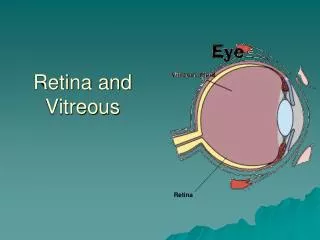
Vitreous and RetinaPediatric Ophthalmology Juan G. Santiago, MD
Hereditary Macular Dystrophies • Stargardt Disease • Fundus flavimaculatus • Best Vitelliform Dystrophy • Familial Drusen
Stargardt Disease • AR or less AD • Juvenile macular degeneration with flecks • Most common • Onset first 2 decades • Symptoms • Decreased vision • Nyctalopia
Stargardt Disease • Findings • Foveal atrophy • Bull’s eye maculopathy • Bilateral pisciform flecks at level of RPE • Change with time • Beaten-bronze fundus • Pathology • RPE massively thickened by accumulation of lipofuscin
Stargardt Disease • FA • Dark choroid (85%) • Hyperfluorescent pisciform flecks • ERG and EOG • Normal or subnormal
Fundus flavimaculatus • AR • Findings • Bilateral pisciform flecks at level of RPE • Predominantly involves peripheral retina • Central vision preserved until macula involved
Fundus flavimaculatus • Pathology • Lipofuscin deposits within hypertrophied RPE cells • ERG • Degree of abnormality correlates with amount of fundus involvement
Best Vitelliform Dystrophy • AD • Progressive with onset in 1st decade of life • RPE is primarily affected • Exudative central macular detachment; SRNVM • Stages • Carrier state • Normal fundus, abnormal EOG • Previtelliform stage • Small round submacular dot • Vitelliform stage • Yellow-orange egg yolk/fried egg appearance; can be multiple; usually between 3-15 yrs
Best Vitelliform Dystrophy • Scrambled egg stage • Irregular subretinal spots; vision usually still good • Cyst stage • Pseudohypopyon stage • RPE atrophy • Round chorioretinal atrophy stage • Atrophic scar • ERG: normal • EOG: abnormal
Familial Drusen • AD • Small yellow-white, round to oval deposits on Bruch’s membrane • Decreased vision after 40 yrs • Complications • Macular edema • Hemorrhage • SRNVM
Hereditary Vitreoretinopathies • Juvenile Retinoschisis • Stickler Syndrome • Familial Exudative Vitreoretinopathy • Norrie Disease • Goldmann-Favre Vitreoretinal Dystrophy
Juvenile Retinoschisis • X-linked recessive • Cleavage of retina at NFL • Foveal (almost all) • Peripheral (~50%) • Findings • Foveal star-shaped or spokelike configuration • Vitreous veils or strands • Vitreal syneresis
Juvenile Retinoschisis • Complications • Vitreous hemorrhages • Retinal detachments • ERG • Normal a-wave • Reduced b-wave • EOG • Normal
Stickler Syndrome • AD • Ophthalmic manifestations • High myopia • Lattice degeneration • Retinal hole or tears • Retinal detachment • Optic atrophy • Cataract • Glaucoma • Ectopia lentis
Stickler Syndrome • Orofacial abnormalities • Pierre Robin anomaly • Midfacial flattening • Cleft palate • Marfinoid habitus • Hearing loss • Mitral valve prolapse • Joint abnormalities • Hyperextensibility • Enlargement • Arthritis
Familial Exudative Vitreoretinopathy • AD or X-linked recessive • Vitreous traction and PVD • Avascular peripheral retina, white w/o pressure, vitreous bands, straightened vessels • Fibrovascular proliferation with neovascularization, exudates, dragging of disc and macula, retinal folds and detachment
Norrie Disease • X-linked recessive • Defect of retinal development • Findings • Bilateral leukocoria • White, often hemorrhagic retrolental mass • Retinal dysplasia • Peripheral NV • Hemorrhagic RD • Retinal necrosis • Other findings • Deafness • Mental retardation
Goldmann-Favre Vitreoretinal Dystrophy • AR • Decreased vision + nyctalopia • Vitreous strands + veils • Central and peripheral retinoschisis • Optic disc pallor • Attenuated retinal vessels • Nummular pigmetary changes • ERG: markedly reduced • EOG: abnormal
Systemic Diseases and Disorders With Retinal Manifestations • Diabetes Mellitus • Leukemia • Albinism • Familial Oculorenal Syndromes • Cherry-red spot • Gangliosidoses
Prevalence of NPDR Rarely: < 3 yrs onset 50%: >7 yrs onset 90%: >15 yrs onset NPDR result from obstruction of retinal capillaries and abnormal capillary permeability Findings Microaneurysms Retinal hemorrhages Areas of retinal nonperfusion Cotton-wool spots Hard exudates IRMA Venous dilation Insulin Dependent Diabetes Mellitus (Type I)
Insulin Dependent Diabetes Mellitus (Type I) • ↑ Glucose • Myopia • ↓ Glucose • Hyperopia • Diabetics cataracts • Collection of sorbitol within the lens • Surveillance • Poor control Initial visit 9 yrs and annual f/up • Good control Initial visit 3 yrs after puberty and annual f/up
Retinopathy children < adults Poor prognosis Choroid most affected Retinal findings Retinal hemorrhages Flame shaped White centered hemorrhages Leukemic infiltrates vs fibrin thrombi??? Perivascular infiltrations Microinfarction Tumor infiltrations Leukemia
Leukemia • Optic nerve infiltration • Emergent radiation therapy • Anterior segment infiltration • Heterochromia iridis • Iris infiltrates • Pseudohypopyon • Spontaneous hyphema • Glaucoma • Orbit infiltration
Oculocutaneous albinism Tyrosinase-neg (no pigmentation) Tyrosinase-pos (some pigmentation) X-linked ocular albinism Findings Iris transillumination Foveal aplasia / hypoplasia Albinotic (hypopigmented) fundus Nystagmus All forms of albinism are heritable, GENETIC counseling is important!! Albinism
Syndromes Chediak Higashi Susceptible to infections Hermansky Pudlak Bleeding diathesis Puerto Rican descent Izquierdo eMedicine Albinism
Lowe (oculocerebral) syndrome X-linked recessive Non-ocular features Aminoaciduria, proteinuria, hematuria Mental retardation, hypotonia, areflexia Family members Mothers have punctate snowflake opacities Ocular features Cataract + glaucoma Alport syndrome X-linked Nerve deafness Nephritis Anterior lenticonus Familial renal-retinal dystrophy AR Interstitial nephritis Pigmentary retinal degeneration Familial Oculorenal Syndrome
Cherry Red Spot • Loss of transparency of the perifoveal retinal due to edema or deposition of abnormal materials in the retinal ganglion cells. • Most common causes • Tay-Sachs disease • Sandhoff disease • Nieman-Pick disease • Sialidosis • Farber lipogranulomatosis • Metachromic leukodystrophy • GM1 gangliosidosis • CRAO • Trauma (macular edema)
Group of lysosomal lipid storage disorders GM1 gangliosidoses Deficiency of beta-galactosidase Derry disease GM2 gangliosidoses Deficiency of the enzyme hexosaminidase. Tay Sachs disease Sandhoff disease Bernheimer-Seitelberger Gangliosidoses