Download
1 / 11
110 likes | 222 Views
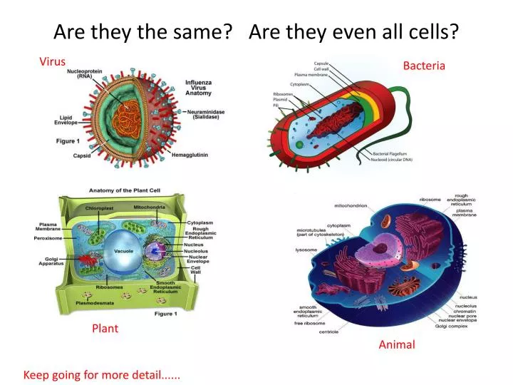
Are they the same? Are they even all cells?. Virus. Bacteria. Plant. Animal. Keep going for more detail. Virus (thingy... Cell?). http://www.chm.bris.ac.uk/webprojects2006/Kelly/influenzafigure1.jpg. Bacteria. http://ilovebacteria.com/Images/bacterialcell.jpg. Plant cell.

E N D
Are they the same? Are they even all cells? Virus Bacteria Plant Animal Keep going for more detail......
Virus (thingy... Cell?) http://www.chm.bris.ac.uk/webprojects2006/Kelly/influenzafigure1.jpg
Bacteria • http://ilovebacteria.com/Images/bacterialcell.jpg
Plant cell • http://www.uvm.edu/~inquiryb/webquest/fa06/mvogenbe/plantcell.jpg
Animal cell • http://www.uvm.edu/~inquiryb/webquest/fa06/mvogenbe/Animal-Cell.jpg
LINKS animation site by John Kyrk • Cell Biology .... Everything and more (lots of animations) ......http://www.johnkyrk.com/ • Cell Anatomy animation..... http://www.johnkyrk.com/CellIndex.html • DNA - Protein Synthesis animation.... http://www.johnkyrk.com/er.html • A WOW timeline of everything (big Bang – now).... http://www.johnkyrk.com/evolution.html
Procedure for observing Plasmolysis in Rhubarb (or red onion): • Remove a v thin slice of rhubarb plant (as red as possible) and place it on a microscope slide. • Use a dropper to place a drop of water on the slice. Cover with a coverslip and place the slide under a microscope. • Focus under 100x power. Move the slide around to find an area where you can clearly see single cells. They will appear rectangular and look like red bricks in a wall. Leave the slide on the microscope. • Using a dropper, place a drop of sugar solution on the right edge of the coverslip. Place a small piece of paper towel on the left edge of the coverslip. As the fresh water soaks into the paper towel it will draw the salt water under the coverslip. This is illustrated below (next slide). • Observe the same cells you were looking at before adding the salt solution. You will notice that the cell contents have shrunken down, leaving a space between the cytoplasm and the cell wall. This is called plasmolysis. • Make a sketch of the appearance of a few of the cells • Repeat Step 4 using fresh water. It may take a number of tries to remove all of the saltwater. When successful, the cells will appear as normal. 8. Clean up the slide and coverslip and return the microscope to its proper place. Questions • In which direction was the water moving (into or out of the cell) during plasmolysis? • What prevents the rhubarb cells from completely collapsing?
Rhubarb plasmolysis setup..... coverslip Sugar solution Paper towel rhubarb Microscope slide
Does is look like this..... • Before (in water)... • After (in sugar solution)...