Download
1 / 34
340 likes | 458 Views
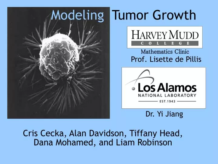
Modeling Tumor Growth. Mathematics Clinic. Prof. Lisette de Pillis. Dr. Yi Jiang. Cris Cecka, Alan Davidson, Tiffany Head, Dana Mohamed, and Liam Robinson. Los Alamos National Lab. Operated by : University of California For : Department of Energy Location : Northern New Mexico

E N D
Modeling Tumor Growth Mathematics Clinic Prof. Lisette de Pillis Dr. Yi Jiang Cris Cecka, Alan Davidson, Tiffany Head, Dana Mohamed, and Liam Robinson
Los Alamos National Lab • Operated by: University of California • For: Department of Energy • Location: Northern New Mexico • Missions: • National Security • Scientific Research
Social Implications • Cancer - the 2nd leading cause of death in the U.S. • Chemotherapy harmful to patient • Better tumor models can help to develop more effective treatments
Given: • Model of a tumor spheroid • No blood vessels • Very small • Goals: • To extend model to include blood vessels • Different vasculature structures • To study chemotherapy treatments Scanning Electron Micrograph The Main Goal *http://www.vet.purdue.edu/cristal/sem-spheroid1-black.gif
Model Description • The three cell types within the model are: • proliferating cells: alive, can divide and grow • quiescent cells: alive, but dormant • necrotic cells: dead
Tumor described on 3 biological levels: • Cellular: • 3D grid of ‘sites’ created • Cells can grow and occupy multiple sites • Extracellular: • Nutrients, waste, chemicals diffuse through tumor cells • Subcellular: • Chemical concentrations cause the cells to respond Model Description
Simulated Model Cross-Section • Grid site • Tumor cell
Initialize Monte Carlo Movement Solve Diffusion Equation Determine Protein Expression Chemicals/Volume Favorable? Quiescent/Necrotic Time to Divide? Possible Cell Shedding if on Surface Divide into 2 Cells *Adapted from a flow chart in: Yi Jiang et. al. “A Multiscale Model for Avascular Tumor Growth”
Monte Carlo • A stochastic algorithm • Strategy • Make a random change • Find a border • Change cell ownership • Calculate the difference in energy • Accept/Reject change • Boltzmann factor
Chemical Diffusion • Chemicals the cells use in this model: • O2, Glucose, Waste, Growth Factors, and Inhibitory Factors • Modeling the time-dependent chemical diffusion equation: • Finite Difference Approximations • yields a linear system of equations
E2F Cell Cycle Proliferating Cells Quiescent Cells GSK3b TGFb SCF SMAD P15 P27 P21 CyCD, CDK4 CyCE, CDK2 Rb S phase *Adapted from a flow chart in Yi Jiang et. al. “A Multiscale Model for Avascular Tumor Growth” *www.bmb.psu.edu/courses/biotc/489/biointeract.htm
Addition of Vasculature • New blood vessel ‘cell’ type added: • can occupy sites • constant chemical concentrations • Reasonable as the speed of the relevant chemical diffusion is slow compared to the rate of blood flow through the vessel.
Vasculature Structure • Can select one of three different vasculature structures • Single Vein • Grid Lattice Structure • Hexagonal Lattice Structure • Have been observed in biological tumors • Adds a greater degree of flexibility to the model • Allows for more structural options to be added later
Extending the Monte Carlo • Extend the J-matrix to include vasculature • Vasculature should be static • Other cells should not encroach upon vasculature • The vasculature should not grow
PDE Solver • Recall the time-dependent diffusion equation: • To solve with arbitrary boundary conditions • We use a Backwards Euler approximation • Stable Linear System • Solve linear system with Gauss-Seidel method • Iterative method • Stable, guaranteed convergence for our system • Strictly (but weakly) Diagonally Dominant
Vasculature and BCs • Treat vasculature as boundary conditions • Can be in an arbitrary geometry • PDE solver supports this applying the identity iteration.
Grid Sites 200 Grid Sites 200 0 Grid Sites 200 0 Grid Sites 200 Avascular vs Vascularized Tumor No Vasculature Constant Line Vasculature
Grid Sites 200 Grid Sites 200 0 Grid Sites 200 0 Grid Sites 200 Delayed vs Constant Vasculature Line Delayed Constant
Grid Sites 200 Grid Sites 200 0 Grid Sites 200 0 Grid Sites 200 Delayed vs Constant Vasculature Square Grid Delayed Constant
Grid Sites 200 Grid Sites 200 0 Grid Sites 200 0 Grid Sites 200 Delayed vs Constant Vasculature Hexagonal Grid Delayed Constant
Types of Chemotherapeutic Agents • Cell Cycle Specific vs. Non Cell Cycle Specific • Alkylating Agents • Nitrosoureas • Antimetabolites • Anthracyclines • Topoisomerase II Inhibitors • Mitotic Inhibitors • Corticosteroid Hormones
Apoptosis vs. Necrosis • Two types of cell death: • Apoptosis • Necrosis While necrosis leaves debris after cell death occurs, apoptosis does not. This has implications for the diffusion of chemicals.
Drug Pharmacokinetics • Cancerboard.ab.ca, www.Canceractive.com • Route of administration • Dose administered • Dosing interval • Plasma drug concentrations
Modeling Chemotherapy • Added cyclophosphamide as a new chemical • Regularly scheduled doses once the vasculature is created • Blood plasma concentration • Constant boundary condition during each step • Changes from step to step to simulate AUC profile • Stochastic model determines if cells become apoptotic based on drug concentration • Apoptotic cells replaced by medium
Limitations of the Model for Chemo • Hardware constraints • Patient toxicity • Chemotherapy drug cocktails
Grid Sites 200 Grid Sites 200 Grid Sites 200 0 Grid Sites 200 0 Grid Sites 200 0 Grid Sites 200 Chemotherapy Treatments 37 MCS Low Dose Chemotherapy High Dose Chemotherapy No Treatment
Grid Sites 200 Grid Sites 200 Grid Sites 200 0 Grid Sites 200 0 Grid Sites 200 0 Grid Sites 200 Chemotherapy Treatments 40 MCS Low Dose Chemotherapy High Dose Chemotherapy No Treatment
Grid Sites 200 Grid Sites 200 Grid Sites 200 0 Grid Sites 200 0 Grid Sites 200 0 Grid Sites 200 Chemotherapy Treatments 50 MCS Low Dose Chemotherapy High Dose Chemotherapy No Treatment
Grid Sites 200 Grid Sites 200 Grid Sites 200 0 Grid Sites 200 0 Grid Sites 200 0 Grid Sites 200 Chemotherapy Treatments 60 MCS Low Dose Chemotherapy High Dose Chemotherapy No Treatment
Grid Sites 200 Grid Sites 200 Grid Sites 200 0 Grid Sites 200 0 Grid Sites 200 0 Grid Sites 200 Chemotherapy Treatments 64 MCS Low Dose Chemotherapy High Dose Chemotherapy No Treatment
Chemotherapy Treatments Low Dose Chemotherapy High Dose Chemotherapy No Treatment
Future Work • Chemotherapy experiments that allow the tumor to reach a detectable size • Inclusion of multiple chemotherapy drugs, including cell cycle specific varieties • Patient toxicity simulation • Optimal control • Treatment schedule • Dose level
Acknowledgments • Prof. Lisette DePillis, Advisor • Dr. Yi Jiang, Liason • Prof. Michael Raugh, Clinic Director • Los Alamos National Lab, Sponsor • Barbara Schade, Administrative Assistant • Claire Connelly, System Administrator